
Rabbit anti-LAIR1 Recombinant Monoclonal Antibody(S-285-2)别名宿主反应种属应用免疫原形式浓度纯化方法类型克隆号同种型储存/保存方法存储溶液背景说明细胞定位UniProt
| 概述 | |
| 别名 |
Leukocyte-associated immunoglobulin-like receptor 1; LAIR-1; hLAIR1; CD305
|
| 宿主 |
Rabbit
|
| 反应种属 |
Human
|
| 应用 |
WB: 1:1000, IP: 1: 50, IHC-P: 1:500, ICC: 1:500, FC(Extra): 1:500
|
| 免疫原 |
Recombinant protein
|
| 性能 | |
| 形式 |
Liquid
|
| 浓度 |
0.5 mg/mL
|
| 纯化方法 |
Protein A affinity column
|
| 类型 |
Monoclonal Antibody
|
| 克隆号 |
S-285-2
|
| 同种型 |
IgG
|
| 储存/保存方法 |
Store at -20℃ for one year.
|
| 存储溶液 |
PBS, 40% Glycerol, 0.05% BSA, 0.03% Proclin 300
|
| 靶标 | |
| 背景说明 |
Leukocyte-associated immunoglobulin-like receptor 1 is a protein that in humans is encoded by the LAIR1 gene [PMID: 9285412]. LAIR1 has also been designated as CD305 (cluster of differentiation 305). LAIR-1 is a 32 kDa transmembrane glycoprotein with a single immunoglobulin-like domain and a cytoplasmic tail containing two immune receptor tyrosine-based inhibitory motifs. LAIR-1 recruits SHP-1 and SHP-2 phosphatases upon activation, and cross-linking of the LAIR-1 antigen on natural killer (NK) cells results in strong inhibition of NK cell-mediated cytotoxicity [PMID: 9285412].
|
| 细胞定位 |
Cell membrane
|
| UniProt |
Q6GTX8
|
实验结果图
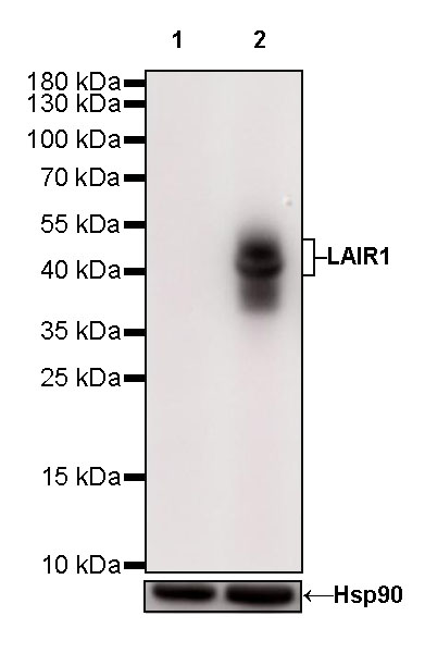
WB result of LAIR1 Rabbit mAb Primary antibody: LAIR1 Rabbit mAb at 1/1000 dilution Lane 1: HeLa whole cell lysate 20 µg Lane 2: THP-1 whole cell lysate 20 µg Negative control: HeLa whole cell lysate Secondary antibody: Goat Anti-Rabbit IgG, (H+L), HRP conjugated at 1/10000 dilution Predicted MW: 31 kDa Observed MW: 40~50 kDa Exposure time: 28 s
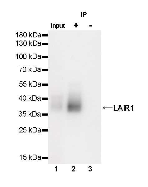
LAIR1 Rabbit mAb at 1/50 dilution (1µg) immunoprecipitating LAIR1 in 0.4 mg THP-1 whole cell lysate. Western blot was performed on the immunoprecipitate using LAIR1 Rabbit mAb at 1/1000 dilution. Secondary antibody (HRP) for IP was used at 1/400 dilution. Lane 1: THP-1 whole cell lysate 20µg (Input) Lane 2: LAIR1 Rabbit mAb IP in THP-1 whole cell lysate Lane 3: Rabbit monoclonal IgG IP in THP-1 whole cell lysate Predicted MW: 40 kDa Observed MW: 40 kDa Exposure time: 150 s

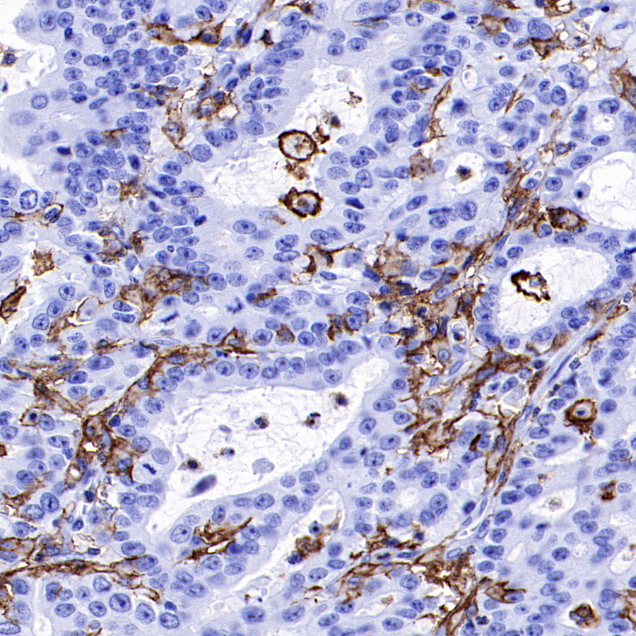
IHC shows positive staining in paraffin-embedded human tonsil. Anti-LAIR1 antibody was used at 1/500 dilution, followed by a HRP Polymer for Mouse & Rabbit IgG (ready to use). Counterstained with hematoxylin. Heat mediated antigen retrieval with Tris/EDTA buffer pH9.0 was performed before commencing with IHC staining protocol.
IHC shows positive staining in paraffin-embedded human stomach. Anti-LAIR1 antibody was used at 1/500 dilution, followed by a HRP Polymer for Mouse & Rabbit IgG (ready to use). Counterstained with hematoxylin. Heat mediated antigen retrieval with Tris/EDTA buffer pH9.0 was performed before commencing with IHC staining protocol.
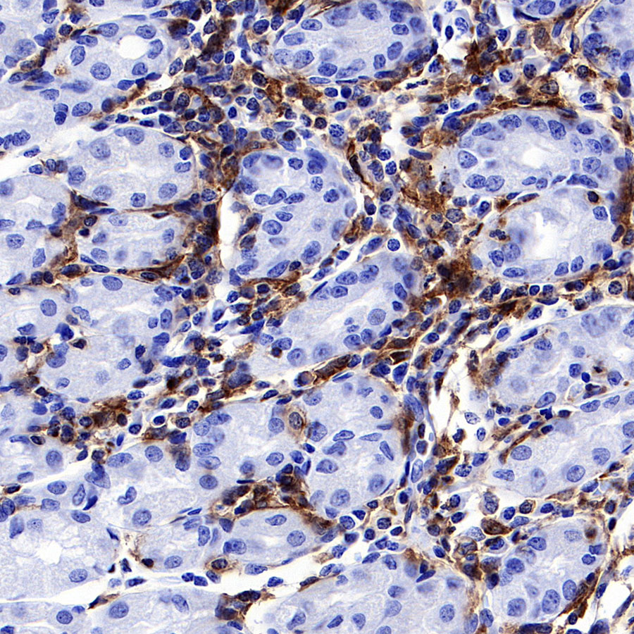
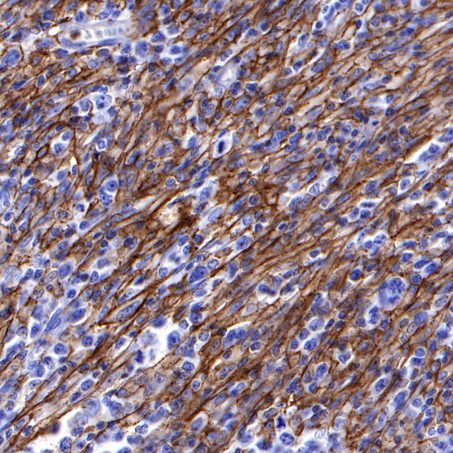
IHC shows positive staining in paraffin-embedded human ovarian cancer. Anti-LAIR1 antibody was used at 1/500 dilution, followed by a HRP Polymer for Mouse & Rabbit IgG (ready to use). Counterstained with hematoxylin. Heat mediated antigen retrieval with Tris/EDTA buffer pH9.0 was performed before commencing with IHC staining protocol.
IHC shows positive staining in paraffin-embedded human gastric cancer. Anti-LAIR1 antibody was used at 1/500 dilution, followed by a HRP Polymer for Mouse & Rabbit IgG (ready to use). Counterstained with hematoxylin. Heat mediated antigen retrieval with Tris/EDTA buffer pH9.0 was performed before commencing with IHC staining protocol.
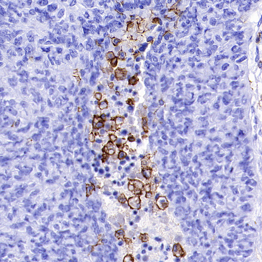
IHC shows positive staining in paraffin-embedded human diffuse large B-cell lymphoma. Anti-LAIR1 antibody was used at 1/500 dilution, followed by a HRP Polymer for Mouse & Rabbit IgG (ready to use). Counterstained with hematoxylin. Heat mediated antigen retrieval with Tris/EDTA buffer pH9.0 was performed before commencing with IHC staining protocol.
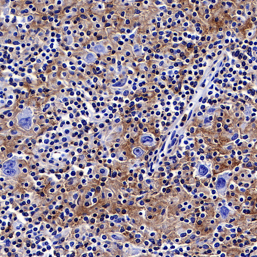
IHC shows positive staining in paraffin-embedded human Hodgkin’s lymphoma. Anti-LAIR1 antibody was used at 1/500 dilution, followed by a HRP Polymer for Mouse & Rabbit IgG (ready to use). Counterstained with hematoxylin. Heat mediated antigen retrieval with Tris/EDTA buffer pH9.0 was performed before commencing with IHC staining protocol.
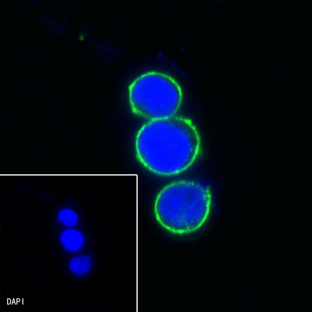
ICC shows positive staining in THP-1 cells. Anti-LAIR1 antibody was used at 1/500 dilution (Green) and incubated overnight at 4°C. Goat polyclonal Antibody to Rabbit IgG – H&L (Alexa Fluor® 488) was used as secondary antibody at 1/1000 dilution. The cells were fixed with 100% ice-cold methanol and permeabilized with 0.1% PBS-Triton X-100. Nuclei were counterstained with DAPI (Blue).
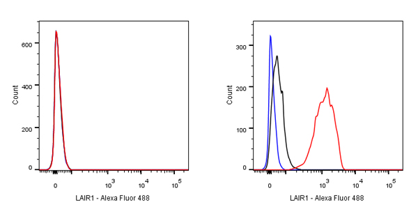
Flow cytometric analysis of HeLa (Human cervix adenocarcinoma epithelial cell, left) / THP-1 (Human monocytic leukemia monocyte, right) cells labelling LAIR1 antibody at 1/500 dilution (0.1 μg) / (red) compared with a Rabbit monoclonal IgG (Black) isotype control and an unlabelled control (cells without incubation with primary antibody and secondary antibody) (Blue). Goat Anti-Rabbit IgG Alexa Fluor 488 was used as the secondary antibody. Negative control: HeLa