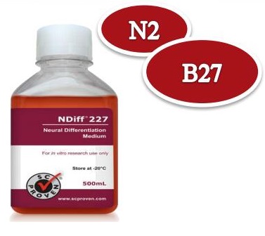
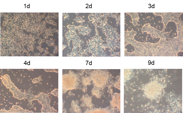
2)这个实验中,干细胞不形成聚落,单层生长。这个实验中外源因子很少,高度可控。因此,这个实验成为了体外诱导分化干细胞成为神经细胞的标准实验方案。这个培养基,就是Cellartis的NDiff 227培养基。(conversion of embryonic stem cells into neuroectodermal precursors in adherent monoculture)。
Estrogen-regulated protein LIV-1; Solute carrier family 39 member 6; Zrt- and Irt-like protein 6; ZIP-6; Zinc transporter ZIP6
宿主
Rabbit
反应种属
Human
应用
WB: 1:1000, IHC-P: 1:200, ICC: 1:250
免疫原
Synthetic peptide
性能
形式
liquid
浓度
0.5 mg/ml
纯化方法
Protein A affinity column
类型
Monoclonal antibody
克隆号
206-8224
储存/保存方法
Store at -20℃ for one year.
存储溶液
PBS, 40% Glycerol, 0.05% BSA, 0.03% Proclin 300
靶标
背景说明
Zinc transporter LIV-1 (SLC39A6) is estrogen regulated and present in increased amounts in estrogen receptor-positive breast cancer as well as in tumors that spread to the lymph nodes. The LIV-1 subfamily of ZIP zinc transporters consists of nine human sequences that share considerable homology across transmembrane domains. Many of these sequences have been shown to transport zinc and/or other ions across cell membranes. SLC39A6 is a member of the ZIP family of transporters, which control zinc homeostasis by regulating the influx of zinc from extracellular to intracellular spaces. The zinc transport function of SLC39A6 plays an important role in cellular metabolism. Zinc is required for a variety of cellular processes, including immune activity, protein synthesis, nucleic acid metabolism, cell proliferation, tissue repair and cell division, and low zinc levels can lead to metabolic disorder and inhibit cell growth. Overexpression of SLC39A6 has been related to the progression of several types of cancer, including breast , prostate, pancreatic, cervical and liver cancer, verexpression of SLC39A6 promoted the epithelial-mesenchymal transition (EMT).
细胞定位
Membrane, cytoplasm
UniProt
Q13433
实验结果图
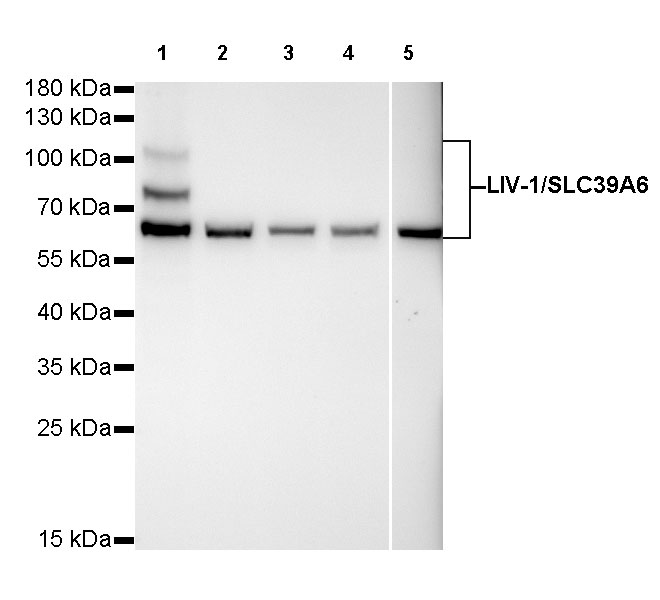
WB result of LIV-1/SLC39A6 Rabbit mAb Primary antibody: LIV-1/SLC39A6 Rabbit mAb at 1/1000 dilution Lane 1: MCF7 whole cell lysate 20 µg Lane 2: MDA-MB-231 whole cell lysate 20 µg Lane 3: Hela whole cell lysate 20 µg Lane 4: HepG2 whole cell lysate 20 µg Lane 5: 293T whole cell lysate 20 µg Secondary antibody: #JP20040 at 1/10000 dilution Predicted MW: 85 kDa Observed MW: 103/85/68 kDa Exposure time: 120s(Ultra High Sensitivity ECL)
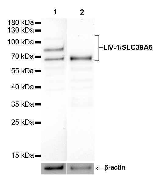
WB result of LIV-1/SLC39A6 Rabbit mAb Primary antibody: LIV-1/SLC39A6 Rabbit mAb at 1/1000 dilution Lane 1: MCF7 whole cell lysate 16 µg Lane 2: MCF7 whole cell lysate (deglycosylated with PNGase F) 16 μg Secondary antibody: #JP20040 at 1/10000 dilution Predicted MW: 85 kDa Observed MW: 103/85/68 kDa Exposure time: 180s
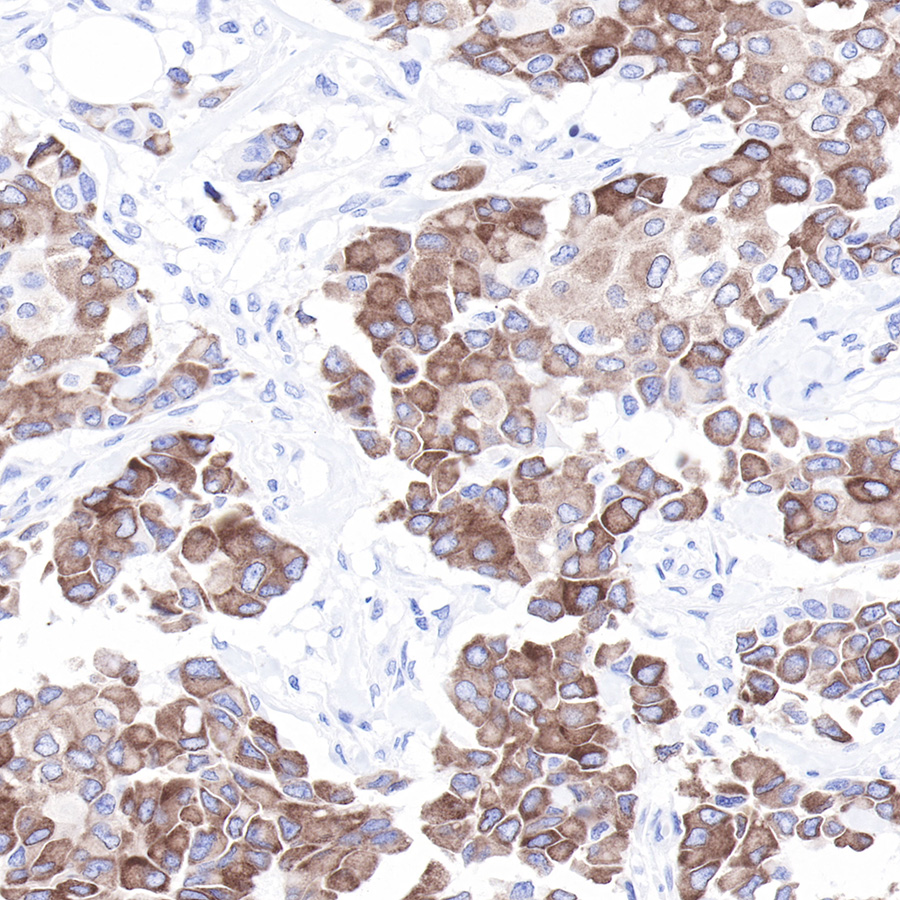
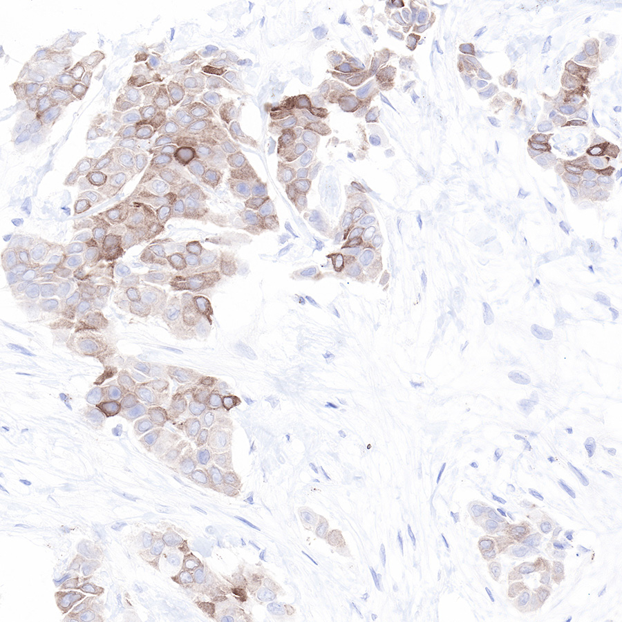
IHC shows positive staining in paraffin-embedded human breast cancer. Anti-LIV-1/SLC39A6 antibody was used at 1/200 dilution, Secondary antibody: #JP20040. Counterstained with hematoxylin. Heat mediated antigen retrieval with Tris/EDTA buffer pH9.0 was performed before commencing with IHC staining protocol.
IHC shows positive staining in paraffin-embedded human breast cancer. Anti-LIV-1/SLC39A6 antibody was used at 1/200 dilution, Secondary antibody: #JP20040. Counterstained with hematoxylin. Heat mediated antigen retrieval with Tris/EDTA buffer pH9.0 was performed before commencing with IHC staining protocol.
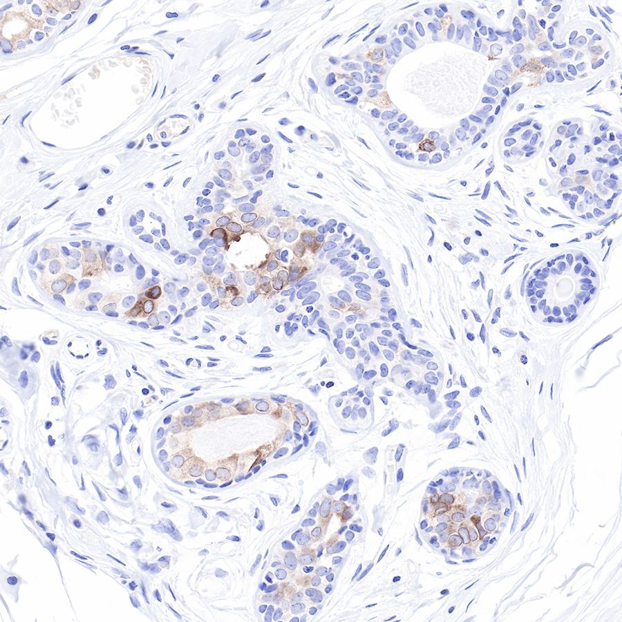
IHC shows positive staining in paraffin-embedded human breast. Anti-LIV-1/SLC39A6 antibody was used at 1/200 dilution, Secondary antibody: #JP20040. Counterstained with hematoxylin. Heat mediated antigen retrieval with Tris/EDTA buffer pH9.0 was performed before commencing with IHC staining protocol.
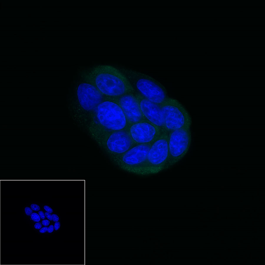
ICC shows positive cytoplasm staining in MCF7 cells. Anti-LIV-1/SLC39A6 antibody was used at 1/250 dilution and incubated overnight at 4°C. Secondary antibody: #JP20025 at 1/1000 dilution. The cells were fixed with 4% PFA and permeabilized with 0.1% PBS-Triton X-100. Nuclei were countersained with DAPI.