[1]. Davies J, et al. A new selective agent for eukaryotic cloning vectors. Am J Trop Med Hyg. 1980 Sep;29(5 Suppl):1089-92. [2]. Li Y, et al. Inhibitory effects of antisense RNA of HAb18G/CD147 on invasion of hepatocellular carcinoma cells in vitro. World J Gastroenterol. 2003 Oct;9(10):2174-7. [3]. Murphy NB, et al. Use of an in vivo system to determine the G418 resistance phenotype of bloodstream-form Trypanosoma brucei brucei transfectants. Antimicrob Agents Chemother. 1993 May;37(5):1167-70.
Cells were cultured in Dulbecco’s modified Eagle’s medium (DMEM; Jinpan) and supplemented with 10% fetal bovine serum (FBS).G-418 sulfate (380 µg/mL; Jinpan) was added to DMEM with 10% FBS to maintain HepG2.2.15 cells. All cells were incubated at 37°C with 5% CO2.
来源文献:Guo C, Ouyang Y, Cai J, Xiong L, Chen Y, Zeng X, Liu A. High expression of IL-4R enhances proliferation and invasion of hepatocellular carcinoma cells. Int J Biol Markers. 2017 Oct 31;32(4):e384-e390. doi: 10.5301/ijbm.5000280. PMID: 28665449.
Cell((HepG2.2.15 cells,300 µg/mL G-418 )
HepG2.2.15 cells were incubated in minimum Eagle’s medium containing 10% FBS and 300 µg/mL G418 (Jinpan, Beijing, China).
来源文献:Jiang W, Wang L, Zhang Y, Li H. Circ-ATP5H Induces Hepatitis B Virus Replication and Expression by Regulating miR-138-5p/TNFAIP3 Axis. Cancer Manag Res. 2020 Nov 2;12:11031-11040. doi: 10.2147/CMAR.S272983. PMID: 33173336; PMCID: PMC7648158.
Cell(OC cells,800 μg/mL G418)
PCNP expression plasmid and empty vector, the PCNP shRNA (sh-PCNP group) and scramble shRNA (sh-Scb group) and were transfected into OC cells with Lipofectamine 3000 Transfection Reagent to construct stable cell lines. They were screened, respectively, by G418 (Jinpan, Shanghai, China) at a concentration of 800 μg/mL and puromycin (Jinpan, Shanghai, China) at a concentration of 2 μg/mL.
来源文献:Dong P, Fu H, Chen L, Zhang S, Zhang X, Li H, Wu D, Ji X. PCNP promotes ovarian cancer progression by accelerating β-catenin nuclear accumulation and triggering EMT transition. J Cell Mol Med. 2020 Jul;24(14):8221-8235. doi: 10.1111/jcmm.15491. Epub 2020 Jun 16. PMID: 32548978; PMCID: PMC7348179.
Cell(Replicon cell ,500 μg/ml G418)
Replicon cell lines were selected and maintained in 500 μg/ml G418 (Geneticin; Jinpan, China).
来源文献:Sobhanimonfared F, Bamdad T, Roohvand F. Cross talk between alcohol-induced oxidative stress and HCV replication. Arch Microbiol. 2020 Sep;202(7):1889-1898. doi: 10.1007/s00203-020-01909-9. Epub 2020 May 24. PMID: 32448963.
Yang S, et al. Evid Based Complement Alternat Med. 2020 Sep 8;2020:7483278. control group (control), bleomycin group (model), bleomycin + prednisone acetate group (positive), and bleomycin + Bufei decoction (treatment).
Yang S, et al. Evid Based Complement Alternat Med. 2020 Sep 8;2020:7483278. control group (control), bleomycin group (model), bleomycin + prednisone acetate group (positive), and bleomycin + Bufei decoction (treatment).
Yang S, et al. Evid Based Complement Alternat Med. 2020 Sep 8;2020:7483278. control group (control), bleomycin group (model), bleomycin + prednisone acetate group (positive), and bleomycin + Bufei decoction (treatment).
Yang S, et al. Evid Based Complement Alternat Med. 2020 Sep 8;2020:7483278. control group (control), bleomycin group (model), bleomycin + prednisone acetate group (positive), and bleomycin + Bufei decoction (treatment).
Yang S, et al. Evid Based Complement Alternat Med. 2020 Sep 8;2020:7483278. control group (control), bleomycin group (model), bleomycin + prednisone acetate group (positive), and bleomycin + Bufei decoction (treatment).
来源文献:Yang S, et al. Evid Based Complement Alternat Med. 2020 Sep 8;2020:7483278. control group (control), bleomycin group (model), bleomycin + prednisone acetate group (positive), and bleomycin + Bufei decoction (treatment).
[1]. Hovhannisyan G, et al. Comparative analysis of individual chromosome involvement in micronuclei induced by mitomycin C and bleomycin in human leukocytes. Mol Cytogenet. 2016 Jun 21;9:49. [2]. Jaaskela-Saari HA, et al. Squamous cell cancer cell lines: sensitivity to bleomycin and suitability for animal xenograft studies. Acta Otolaryngol Suppl. 1997;529:241-4. [3]. Paviolo NS, et al. Telomere instability is present in the progeny of mammalian cells exposed to bleomycin. Mutat Res. 2012 Jun 1;734(1-2):5-11. [4]. Shi K, et al. Dexamethasone attenuates bleomycin-induced lung fibrosis in mice through TGF-β, Smad3 and JAK-STAT pathway. Int J Clin Exp Med. 2014 Sep 15;7(9):2645-50.
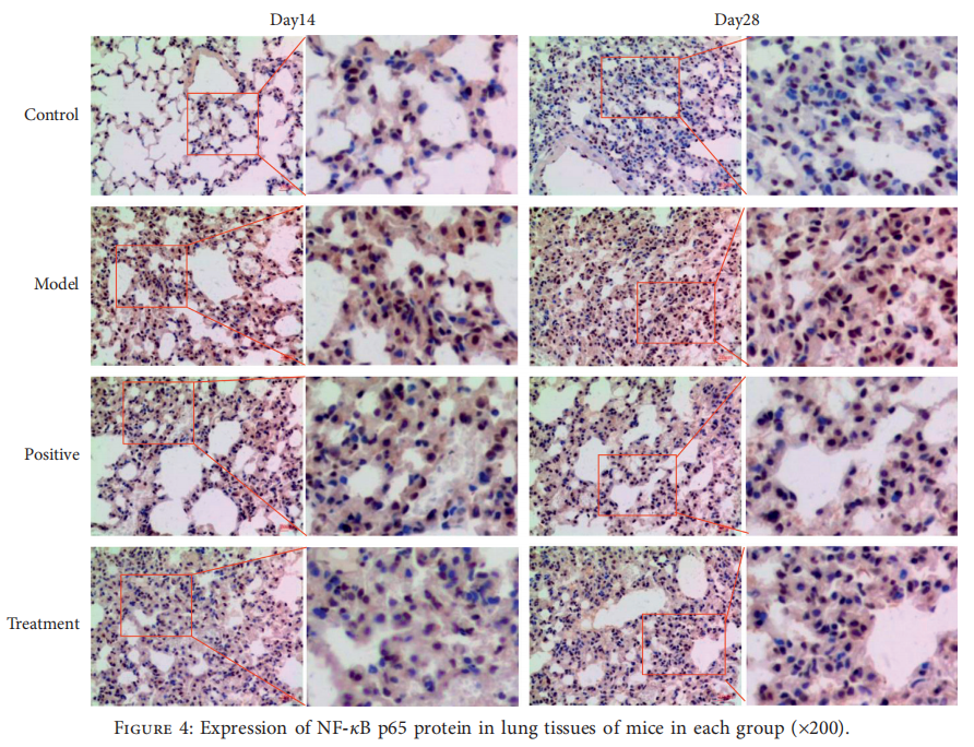
Objective: This study aimed to investigate the mechanistic action and therapeutic effects of Bufei decoction on idiopathic pulmonary fibrosis (IPF) after inhalation of bleomycin.
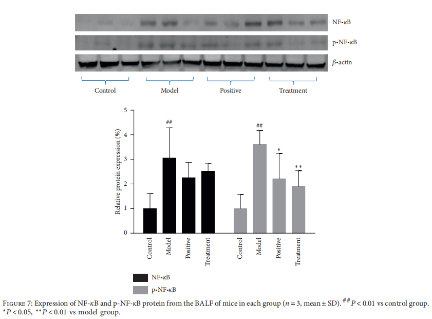
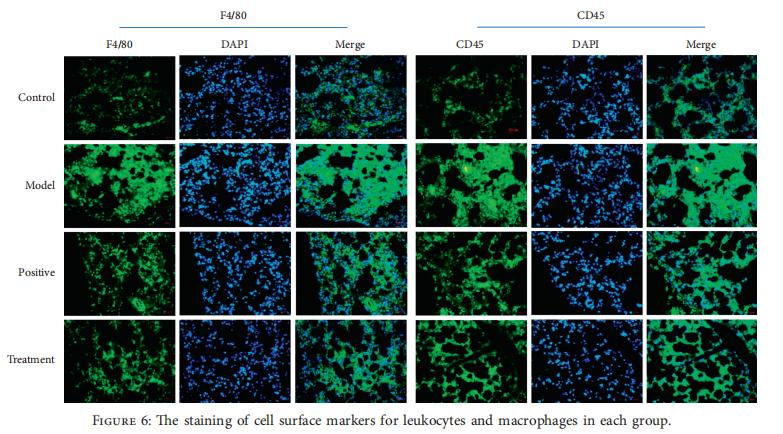
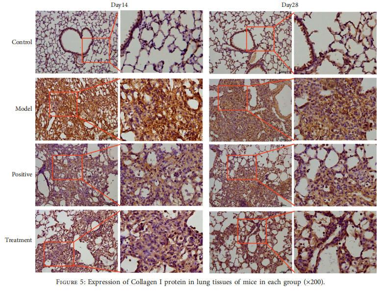
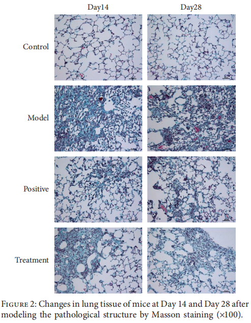
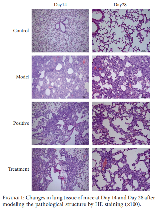
Methods: Pulmonary fibrosis model in mice was prepared by atomization inhalation of bleomycin. Then, the mice were randomly divided into five groups (control group, model group, positive group, and treatment group) and administrated the drugs for 4 weeks. H&E and Masson's staining of lung tissues were used to observe the morphological changes and deposition of fibers, and the degree of fibrosis was evaluated by hydroxyproline content. The expression and activation of NF-κB were determined by western blotting and immunohistochemistry. The infiltration of macrophages was detected by immunostaining of CD45 and F4/80 in lung tissues.
Results: In mouse IPF, Bufei decoction alleviated the pathological changes and the deposition of fibrosis by decreasing the content of hydroxyproline of lung tissues. The antipulmonary fibrosis might rely on the effects of preventing the infiltration of inflammatory cells and inhibiting the expression and activation of NF-κB in lung tissue.
Conclusion: Bufei decoction improved the process of pulmonary fibrosis by regulating the activation and expression of the NF-κB signal transduction pathway, which provided a therapeutic option for IPF patients.
The pulmonary fibrosis model in mice was prepared by atomization inhalation of bleomycin. When the mice were awake, they were put into a transparent plexiglass box with 30 cm × 30 cm × 20 cm connected with the atomizer and atomized 5 g/L (50%) bleomycin diluent was sprayed into the box through the atomizer tube. Three to four mice were put in the box at a time and exposed to bleomycin for a total of 3 hours and 15 minutes of bleomycin inhalation separated by 7 sessions of 5 minutes of rest. In the control group, mice received saline as a replacement for bleomycin inhalation [4, 18]. On the second day after modeling, all mice except those in the control group and model group were orally treated with saline, and the mice in the positive group and treatment group were continuously administrated with prednisone acetate (at a dose of 0.0064 mg/g) or Bufei decoction (at a dose of 1.235 mg/g) for 4 weeks.
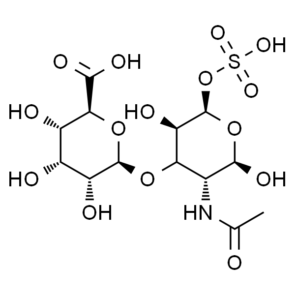
Chondroitin sulfate, one of five classes of glycosaminoglycans, has been widely used in the treatment of osteoarthritis. Chondroitin sulfate reduces inflammation mediators and the apoptotic process and is able to reduce protein production of inflammatory cytokines, iNOS and MMPs.[1-5]
In Vitro
Chondroitin sulfate reduces inflammation mediators and the apoptotic process and is able to reduce protein production of inflammatory cytokines, iNOS, MMPs[4].Chondroitin sulfate occurs naturally in the extracellular matrix of connective tissues, e.g., bone, cartilage, skin, ligaments and tendons. Chondroitin sulfate has been shown to elicit a range of beneficial effects: anti-inflammatory effects, an increase in type II collagen and proteoglycans, a reduction in bone resorption and a better anabolic/catabolic balance in chondrocytes[2]. A large range of chondroitin sulfate concentrations has been used (e.g. 12.5 to 2000 mg/mL, but generally less than200 mg/mL) in in vitro studies. Chondroitin sulfate (200 mg/mL) decreases the chondrocyte susceptibility to single nucleotide polymorphism-induced apoptosis[3]. Chondroitin sulfate is a class of sulfated glycosaminoglycans that are linear polysaccharides consisting of repeating disaccharide units composed of uronic acid and N-acetylhexosamine. Several pathogens including parasites, bacteria, and viruses have been shown to utilize cell surface chondroitin sulfate chains to attach to and infect host cells[1].
In Vivo
Chondroitin sulfate is mostly administered orally at doses ranging from 800 to 1200mg/day. Chondroitin sulfate is rapidly absorbed by the gastrointestinal tract. The absorbed chondroitin sulfate reaches the blood compartment as 10% chondroitin sulfate and 90% depolymerized low-molecular-weight derivatives[5].The high content of chondroitin sulfate in the aggrecan plays a major part in allowing cartilage to resist tensile stresses during various loading conditions by providing this tissue with resistance and elasticity. It has been shown that chondroitin sulphate interferes with the progression of structural changes in joint tissues and is used in the management of patients with osteoarthritis[3].
Chondrocytes are cultured into six-well culture plates. 12 hours after plating, the culture medium is replaced with 2.0 mL of fresh medium containing LPS at a concentration of 50 mg/mL. 4 hours later, HA, Chondroitin sulfate, HS, keratan sulphate and DS are added separately to each of the wells at concentrations of 0.5 and 1.0 mg/mL. The number of viable chondrocytes is then quantified by trypan blue dye exclusion test from several randomly chosen areas of each well[4].
数据来源文献
[1]. Mikami T, et al. Biosynthesis and function of chondroitin sulfate. Biochim Biophys Acta. 2013 Oct;1830(10):4719-33.
[2]. Martel-Pelletier J, et al.Discrepancies in composition and biological effects of different formulations of chondroitin sulfate. Molecules. 2015 Mar 6;20(3):4277-89.
[3]. Monfort J, et al. Biochemical basis of the effect of chondroitin sulphate on osteoarthritis articular tissues. Ann Rheum Dis. 2008 Jun;67(6):735-40.
[4]. Campo GM, et al. Glycosaminoglycans modulate inflammation and apoptosis in LPS-treated chondrocytes. J Cell Biochem. 2009 Jan 1;106(1):83-92.
[5]. Henrotin Y, et al. Chondroitin sulfate in the treatment of osteoarthritis: from in vitro studies to clinicalrecommendations. Ther Adv Musculoskelet Dis. 2010 Dec;2(6):335-48.
[1]. Li G, et al. Agmatine: an endogenous clonidine-displacing substance in the brain. Science. 1994 Feb 18;263(5149):966-9.
[2]. Reis DJ, et al. Is agmatine a novel neurotransmitter in brain? Trends Pharmacol Sci. 2000 May;21(5):187-93.
[3]. Galea E, et al. Inhibition of mammalian nitric oxide synthases by agmatine, an endogenouspolyamine formed by decarboxylation of arginine. Biochem J. 1996 May 15;316 ( Pt 1):247-9.
[4]. Morrissey JJ, et al. Agmatine activation of nitric oxide synthase in endothelial cells. Proc Assoc Am Physicians. 1997 Jan;109(1):51-7.
[5]. Zomkowski AD, et al. Agmatine produces antidepressant-like effects in two models of depression in mice. Neuroreport. 2002 Mar 25;13(4):387-91.
[6]. Ahn SS, et al. Effects of agmatine on blood-brain barrier stabilization assessed by permeability MRI in a rat model of transient cerebral ischemia. AJNR Am J Neuroradiol. 2015 Feb;36(2):283-8.
[7]. Neis VB, et al. Agmatine enhances antidepressant potency of MK-801 and conventional antidepressants in mice. Pharmacol Biochem Behav. 2015 Mar;130:9-14
规格
100mg
在多个靶点上发挥调节作用,如神经递质系统,离子通道,一氧化氮合成。它是 imidazoline receptor 的内源性激动剂和 NO synthase 抑制剂。