醋酸艾塞那肽

醋酸艾塞那肽

货号:
IA2550

品牌:
Jinpan

醋酸艾塞那肽

暂无详情
产品简介
别名 A1155463
英文名称 A-1155463
CAS 1235034-55-5
分子式 C35H32FN5O4S2
分子量 669.79
储存条件 -20°C
纯度 ≥98%
单位
生物活性 A-1155463是一种高度有效、选择性的BCL-XL抑制剂。其对BCL-XL的亲和力属于皮摩尔级别,对BCL-2、BCL-W(Ki=19 nM)、MCL-1(Ki>440 nM)的结合比对BCL-XL弱1000倍以上。[1-3]
In Vitro A-1155463在细胞中破坏BCL-XL-BIM复合体而不是BCL-2-BIM复合体。A-1155463可以杀死BCL-XL依赖性的Molt-4细胞(IC50=70 nM),但对RS4;11细胞没有明显的细胞毒性(EC50>5 mM)。A-1155463在BCL-XL依赖性的H146细胞中诱导凋亡标志,如线粒体释放细胞色素c、caspase的激活、caspase依赖性的sub-G0-G1期DNA的含量积累[2]。
In Vivo 向SCID-Beige不带瘤小鼠中单次腹腔注射5 mg/kg A-1155463,血小板数量在注射后6小时显著下降,在注射后72小时内恢复为正常水平。每天向接种过BCL-XL依赖性H146肿瘤细胞的SCID-Beige小鼠腹腔注射5 mg/kg A-1155463,14天后检测,A-1155463显著抑制了肿瘤的生长,在停止给药后,效果减轻[1]。
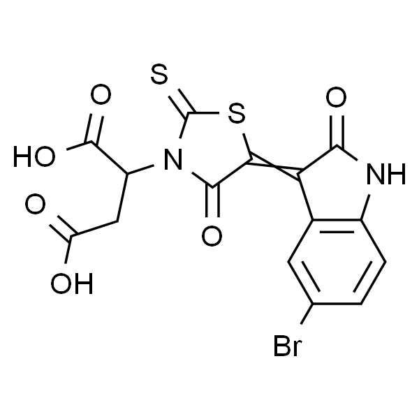
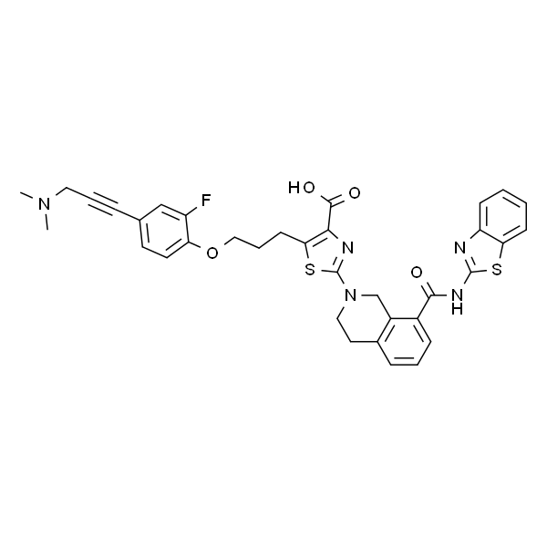
SMILES O=C(C1=C(CCCOC2=CC=C(C#CCN(C)C)C=C2F)SC(N3CC4=C(C=CC=C4C(NC5=NC6=CC=CC=C6S5)=O)CC3)=N1)O
靶点 Bcl-xL
动物实验 Cells are treated with increasing concentration of A-1155463. Cells are assayed for viability after 72 h using the CellTiter-Glo luminescent cell viability assay according to the manufacturer’s protocol. Results are normalized to cells without treatment. EC50 is calculated using the GraphPad Prism software.(Only for Reference) Cell lines: Colorectal cell lines (ATCC)[3]
细胞实验 Animal Models: SCID-Beige小鼠; Dosages: 5 mg/kg ;Administration: 腹腔注射[1]
数据来源文献 [1] Tao ZF, et al. ACS Med Chem Lett. 2014, 5(10):1088-93.
[2] Leverson JD, et al. Sci Transl Med. 2015, 7(279):279ra40.
[3] Haichao Zhang, et al. Molecular Cancer. 2015, 14(1):1-9.
规格 2mg 5mg 10mg

A-1155463是一种高度有效、选择性的BCL-XL抑制剂。

Bcl-w,Rabbit IgG anti-Mouse/Rat/Human,WB/IP/

2cl-w,Rabbit IgG anti-Mouse/Rat/Human,WB/IP/

产品编号: sc-11422     
产品名称: Bcl-w,Rabbit IgG anti-Mouse/Rat/Human,WB/IP/  
供应商: Santa
规格: 200ug
目录价: ¥:2980
库存状态:
CAS编号:
应用范围:
种属来源:
相关信息:
保存条件:
说明书地址: 点击查看详细

引物对ApoPrimer Set (Bcl-2 family)酶试剂盒Takara Clontech

上海金畔生物科技有限公司代理Takara酶试剂盒全线产品,欢迎访问官网了解更多产品信息和订购。

引物对ApoPrimer Set (Bcl-2 family)
品牌 Code No. 产品名称 包装量 价格(元) 说明书 数量
Takara 6623 ApoPrimer Set (Bcl-2 family)(库存销售完毕即终卖) 1 Set ¥1,188 引物对ApoPrimer Set (Bcl-2 family) 引物对ApoPrimer Set (Bcl-2 family) 引物对ApoPrimer Set (Bcl-2 family)
收藏产品 加入购物车
 
■ 制品内容 (for 40 reactions per primer mix)
human Mcl-1 primer mix (each 10 pmol/μl) 40 μl
human Bfl-1 primer mix (each 10 pmol/μl) 40 μl
human Bax-α primer mix (each 10 pmol/μl) 40 μl
human Bcl-2 primer mix (each 10 pmol/μl) 40 μl
human Bak primer mix (each 10 pmol/μl) 40 μl
human Bik primer mix (each 10 pmol/μl) 40 μl
human Bcl-x primer mix (each 10 pmol/μl) 40 μl
human β-actin primer mix (each 10 pmol/μl) 40 μl
APO Positive Control RNA 1 (106 copies/μl) 24 μl
 
■ 制品说明
Human Bcl-2家族蛋白是在细胞凋亡过程中起关键性作用的一类蛋白质,本制品包含扩增human Bcl-2 family基因的cDNA的Primer set。可扩增human Bcl-2 family中的7种基因的cDNA (mcl-1,bfl-1,bax-α,bcl-2,bak,bik,bcl-x*) 和β-actin基因。 (*human Bcl-x primer mix是在bcl-xLbcl-xS基因的相同序列上设计的引物,因此使用同一个引物可分别扩增bcl-xLbcl-xS基因) 。
APO Positive Control 1 RNA中包含上述8种引物的序列,可以与试剂盒中所有的引物进行PCR反应。以Positive Control RNA为模板的扩增产物,与实际mRNA扩增产物的片段大小不同,易于区别。
 
每种引物对扩增产物大小
Primer mix Positive Control RNA来源 mRNA来源
mcl-1 primer mix 358 bp 449 bp
Bfl-1 primer mix 331 bp 413 bp
Bax-α primer mix 330 bp 412 bp
Bcl-2 primer mix 304 bp 380 bp
Bak primer mix 297 bp 371 bp
Bik primer mix 296 bp 370 bp
Bcl-x primer mix 272 bp xL:340 bp
xS
:151 bp
β-actin primer mix 340 bp 275 bp
*:Bcl-x primer mix是在bcl-xLbcl-xS基因的相同序列上设计的引物,因此使用同一个引物可分别扩增bcl-xLbcl-xS基因。
 
■ 保存
-20℃。


页面更新:2024-04-01 15:31:10

A-1331852

A-1331852

货号:
IA2530

品牌:
Jinpan

A-1331852

暂无详情
产品简介
别名 BCL-XL抑制剂(A-1331852)
英文名称 A-1331852
CAS 1430844-80-6
分子式 C38H38N6O3S
分子量 658.81
储存条件 -20°C
纯度 ≥98%
单位
SMILES O=C(C1=NC(N2CC3=C(C=CC=C3C(NC4=NC5=CC=CC=C5S4)=O)CC2)=CC=C1C6=C(C)N(CC7(C8)CC9CC8CC(C9)C7)N=C6)O
靶点 Bcl-xL
规格 5mg 10mg
A-1331852是一种有效的、选择性的BCL-XL抑制剂。

Bcl-XS/L,Rabbit IgG anti-Mouse/Rat/Human,WB/IP/

2cl-XS/L,Rabbit IgG anti-Mouse/Rat/Human,WB/IP/

产品编号: sc-1041     
产品名称: Bcl-XS/L,Rabbit IgG anti-Mouse/Rat/Human,WB/IP/  
供应商: Santa
规格: 200ug
目录价: ¥:2980
库存状态:
CAS编号:
应用范围:
种属来源:
相关信息:
保存条件:
说明书地址: 点击查看详细

Gambogic Acid 藤黄酸

Gambogic Acid 藤黄酸

货号:
IG0050

品牌:
Jinpan

Gambogic Acid  藤黄酸

暂无详情
产品简介
MDL MFCD16878985
别名 Beta-Guttiferrin; ? Guttatic Acid
CAS 2752-65-0
分子式 C38H44O8
分子量 628.75
纯度 HPLC≥98%
单位
生物活性 Gambogic acid 来自 Garcinia hanburyi 树的藤黄树脂。Gambogic acid 抑制 Bcl-XL,Bcl-2,Bcl-W,Bcl-B,Bfl-1 和 Mcl-1,IC50 分别为 1.47 μM,1.21 μM,2.02 μM,0.66 μM,1.06 μM 和 0.79 μM。[1-2]
IC50 Bcl-B:0.66 μM; Mcl-1:0.79 μM; Bfl-1:1.06 μM; Bcl-2:1.21 μM ; Bcl-xL:1.47 μM ; Bcl-W:2.02 μM [1-2]
In Vitro 藤黄酸(Gambogic Acid)是一种药用化合物,来自树木的藤黄树(Garcinia hanburyi)。藤黄酸已经记录了培养中针对肿瘤细胞系的细胞毒活性,其中杀死50%细胞所需的浓度(LD50为~1μM)。使用FPA对抗藤黄酸对6种人抗凋亡Bcl-2家族蛋白的活性。藤黄酸取代不同程度的FITC-BH3肽与所有6种蛋白结合,Bcl-XL的IC50为1.47μM,Bcl-2为1.21μM,Bcl-W为2.02μM,Bcl-B为0.66μM,1.06μM为Bfl-1,Mcl-1为[0.79μM] [1]。在暴露48小时后通过MTT测定评估藤黄酸(GA)或顺铂(CDDP)对A549,NCI-H460和NCI-H1299细胞的生长抑制作用。用Gambogic Acid和CDDP观察到细胞生长的浓度依赖性抑制,A549细胞的IC50为3.56±0.36和21.88±3.21μM,NCI-H460细胞的IC50为4.05±0.51和25.76±4.03μM,和1.12±0.31μM NCI-H1299细胞为25.21±4.38μM[2]。
In Vivo 为了进一步研究藤黄酸是否协同CDDP对抗体内肿瘤生长,将A549肿瘤植入SCID小鼠中。当用CDDP联合藤黄酸处理小鼠时,肿瘤抑制率为69.3%,而单独用CDDP和GA处理的小鼠的肿瘤抑制率分别为57.2%和29.0%[2]。
SMILES O=C(O)/C(C)=CC[C@@]1(C2=O)OC(C)(C)[C@@](C[C@@]2([H])C=C34)([H])[C@]31OC5=C(C(O)=C(C=C[C@](CC/C=C(C)/C)(C)O6)C6=C5C/C=C(C)C)C4=O
靶点 Bcl-2 Family
动物实验 小鼠[2]为了确定藤黄酸与CDDP结合的体内抗肿瘤活性,将活A549细胞(每只小鼠5×10⁶ /100μLPBS)皮下注射到7-8周龄雄性SCID的右侧腹部。老鼠。当平均肿瘤体积达到100 mm3时,将小鼠随机分为4个治疗组,包括对照组(仅生理盐水,n = 5),藤黄酸(每2天3.0 mg/kg,静脉注射; n = 6),CDDP(每周4mg/kg,静脉内; n = 6),和顺序组合(在藤黄酸处理前一天进行CDDP处理,n = 6)。 CDDP(4mg/kg,每周)通常以小于最大耐受剂量的剂量施用,以试图允许更容易检测与铂基试剂和藤黄酸的组合治疗的任何累加效应。用calipre每2天测量一次肿瘤大小。每2天记录一次体重。 14天后,杀死小鼠并切除肿瘤并储存在-80℃直至进一步分析。
细胞实验 通过MTT测定确定藤黄酸,单独的CDDP或组合处理的体外细胞活力效应。将细胞(2×10 4个细胞/ mL)接种到96孔培养板中。孵育过夜后,用藤黄酸(0.44,0.88,1.75,3.5,7,10.5和14μM)处理A549细胞;用藤黄酸(0.5,1,2,4,8,12和16μM)处理NCI-H460细胞;用藤黄酸(0.125,0.25,0.5,1,2和4μM)处理NCI-H1299细胞。对于在NSCLC细胞中的组合治疗,测试三种序列:(a)将藤黄酸随后将CDDP细胞暴露于藤黄酸48小时,然后在冲洗出藤黄酸后,用CDDP处理细胞另外48小时; (b)将CDDP接着将藤黄酸细胞暴露于CDDP 48小时,然后在冲洗掉CDDP后,将细胞用藤黄酸处理另外48小时; (c)同时处理细胞暴露于藤黄酸和ADM两小时48小时。通过使用组合指数(CI)[2]分析药物相互作用的性质。
数据来源文献 [1]. Zhai D, et al. Gambogic acid is an antagonist of antiapoptotic Bcl-2 family proteins. Mol Cancer Ther. 2008 Jun;7(6):1639-46.

[2]. Wang LH, et al. Gambogic acid synergistically potentiates cisplatin-induced apoptosis in non-small-cell lung cancer through suppressing NF-κB and MAPK/HO-1 signalling. Br J Cancer. 2014 Jan 21;110(2):341-52

规格 5mg 10mM*1mL (in DMSO) 10mg 20mg

据报道,可以抑制 Bcl-XL,Bcl-2,Bcl-W,Bcl-B,Bfl-1 和 Mcl-1。

CID5721353, BCL6 inhibitor

CID5721353, BCL6 inhibitorCAS号: 301356-95-6分子式: C15H9BrN2O6S2分子量: 457.27描述储存/保存方法别名外观可溶性/溶解性靶点In vitro(体外研究)In vivo(体内研究)

产品描述
描述

CID5721353 is a B-Cell Lymphoma 6 Inhibitor (BCL6 inhibitor).

储存/保存方法
Store at -20℃ for one year(Powder);Store at 2-4℃ for two weeks;Store at -20℃ for six months after dissolution.
基本信息
别名
CID-5721353; CID 5721353
外观
powder
可溶性/溶解性
DMSO
生物活性
靶点
Bcl-6
In vitro(体外研究)
Small molecular BCL6 inhibitor 79-6 is a cell-permeable oxindole compound that selectively inhibits the transcriptional repression activity of BCL6 but does not affect BCL6 protein levels. 79-6 binds an aromatic pocket situated in the BTB domain lateral groove, induces disruption of BCL6 transcriptional complexes, reactivation of BCL6 target genes and selective killing of BCL6-dependent DLBCL cells. BCL6 can also interact with MTA3 through its RD2 domain and represses the terminal differentiation. It recruits CtBP through its DBD domain and represses its own expression. 79-6 suppresses invasive ability and colony-forming ability in both of MCF-7 and MDA231 cells. The inhibition of BCL6 with 79-6 enhanced the mRNA expression of E-cadherin in both of MCF-7 and MDA231 cells. 79-6 reverses the functions of BCL6 in the positive regulation of EMT, invasion and transcriptional repression of E-cadherin in breast cancer cells.
In vivo(体内研究)
Administration of 50 mg/kg/day 79-6 to SCID mice induced a 65-70% reduction in the size of established BCL6-dependent DLBCL xenografts, but had no effect on BCL6-independent DLBCLs. Therapeutic targeting of BCL6 with the small molecule 79-6 is an effective antilymphoma strategy in vivo.

分子结构图

CID5721353, BCL6 inhibitor

BH3I-1

BH3I-1

货号:
IB1210

品牌:
Jinpan

BH3I-1

暂无详情

BH3I-1

暂无详情
产品简介
MDL MFCD03453544
别名 BHI1
英文名称 BH3I-1
CAS 300817-68-9
分子式 C15H14BrNS2O3
分子量 400.31
储存条件 2-8°C
纯度 ≥97%
单位
生物活性 BH3I-1 is a Bcl-2 family antagonist, which inhibits the binding of the Bak BH3 peptide to Bcl-xL with a Ki of 2.4±0.2 μM in FP assay. BH3I-1 has a Kd of 5.3 μM against the p53/MDM2 pair.[1-3]
In Vitro BH3I-1, while inhibiting its reported target Bcl-2/Bim and Bcl-xL/Bim, shows significant inhibition of both the p53/hDM2 and p300/Hif-1α interactions. This surprising promiscuity, displays by a well studied compound leads to further interrogate the p53/hDM2 interaction utilizing a standard fluorescence polarization (FP) assay with purified protein. The results from the FP assay validates the split-luciferase screen and demonstrates that BH3I-1 has a Kd=5.3 μM against the p53/mDM2 pair, which is comparable to its low micromolar potency reported for the BH3 family of receptors[2]. BH3I-1 inhibits interaction between the BH3 domain and Bcl-xL. NMR analyses reveal that BH3I-1 targets the BH3-binding pocket of Bcl-xL with a Ki of 7.8±0.9 μM[3].
SMILES O=C(O)C(C(C)C)N(C/1=O)C(SC1=CC2=CC=C(Br)C=C2)=S
靶点 Bcl-2 Family;MDM-2/p53
细胞实验 Jurkat cells overexpressing Bcl-xL, FL 5.12 and FL 5.12/Bcl-xL cells (5×10⁴ cells per well) are seeded into white 96-well plates and treated with various concentrations of the compounds (e.g., BH3I-1; 30 μM and 90 μM)for 48 h. For zVAD-FMK protection experiments, cells are preincubated with 100 μM zVAD-FMK for 1 h before the addition of chemicals. Cell viability is determined with an MTS assay with a Victor plate reader. For PI staining experiments, cells are grown in 24-well plates and then incubated with 2 μg/mL PI. Cell death is determined by FACS analysis in a FACSCalibur machine[3].
数据来源文献 [1]. Wang L, et al. Development of dimeric modulators for anti-apoptotic Bcl-2 proteins. Bioorg Med Chem Lett. 2008 Jan 1;18(1):236-40.

[2]. Degterev A, et al. Identification of small-molecule inhibitors of interaction between the BH3 domain and Bcl-xL. Nat Cell Biol. 2001 Feb;3(2):173-82.

[3]. Porter JR, et al. Profiling small molecule inhibitors against helix-receptor interactions: the Bcl-2 family inhibitor BH3I-1 potently inhibits p53/hDM2. Chem Commun (Camb). 2010 Nov 14;46(42):8020-2.

规格 5mg 10mg

BH3I-1是Bcl-XL-BH3 domain interaction抑制剂。它是一种Bcl-2家族蛋白的选择性抑制剂。

CID5721353

CID5721353

货号:
IC2110

品牌:
Jinpan

CID5721353

暂无详情
产品简介
别名 BCL6 inhibitor
英文名称 CID5721353
CAS 301356-95-6
分子式 C15H9BrN2O6S2
分子量 457.28
储存条件 2-8°C
纯度 ≥98%
单位
生物活性 79-6 (CID5721353) (BCL6 inhibitor)是一种BCL6抑制剂,Kd值为138 μM。[3]
In Vitro BCL6抑制剂79-6具有细胞可透过性,可选择性地抑制BCL6的转录抑制活性,但不影响BCL6的蛋白水平[2]。79-6与位于BTB区域侧沟的一个芳香烃口袋结合,诱导BCL6转录复合体被破坏,BCL6靶基因重激活并选择性杀死依赖于BCL6的DLBCL细胞[1]。BCL6通过RD2区域与MTA3相互作用,抑制终末分化。BCL6通过其DBD区域招募CtBP,抑制其自身活性。在MCF-7和MDA231细胞中,79-6抑制细胞入侵能力和集落形成能力,通过抑制BCL6增强E-cadherin的mRNA表达。在乳腺癌细胞中,79-6可逆转BCL6在间充质转化(EMT)正向调节过程、细胞侵染和E-cadherin转录抑制过程中的功能[2]。
In Vivo 对携有BCL6依赖性DLBCL移植瘤的SCID小鼠进行给药(79-6,50 mg/kg/天)可引起期肿瘤尺寸65-70%的减少,但对非BCL6依赖性的DLBCL移植瘤无影响[1]。在体内,79-6抑制BCL6是一种有效的抗淋巴瘤的策略[3]。
SMILES O=C(O)C(N(C/1=O)C(SC1=C2C(NC3=C2C=C(Br)C=C3)=O)=S)CC(O)=O
靶点 Bcl-6 inhibitor
细胞实验 Animal Models: 注射了BCL6依赖性细胞系(OCI-Ly7和SU-DHL6)并形成了肿瘤的SCID小鼠; Dosages: 50 mg/kg; Administration: i.p.[3]
数据来源文献 [1] Cardenas MG, et al. Clin Cancer Res. 2017, 23(4):885-893.
[2] Yu JM, et al. Cancer Lett. 2015, 365(2):190-200.
[3] Sakamoto K, et al. Biochem Biophys Res Commun. 2017, 482
规格 5mg 10mM*1mL in DMSO 10mg 50mg

是一种BCL6抑制剂。

2-乙基-3-甲基戊酰胺

2-乙基-3-甲基戊酰胺

货号:
IA1500

品牌:
Jinpan

2-乙基-3-甲基戊酰胺

暂无详情
产品简介
MDL MFCD12756212
别名 ABT737
英文名称 ABT-737
CAS 852808-04-9
分子式 C42H45ClN6O5S2
分子量 813.43
纯度 ≥98%
单位
生物活性 ABT-737 是一种类似 BH3 的 Bcl-xL, Bcl-2 和 Bcl-w 的抑制剂, EC50 值分别为 78.7 nM, 30.3 nM 和 197.8 nM[1-3]。
IC50 Bcl-2:30.3 nM (EC50)

Bcl-xL:78.7 nM (EC50)

Bcl-W:197.8 nM (EC50)

Bcl-B:1820 nM (EC50) [1-3]

In Vitro ABT-737和ATO以浓度和时间依赖性方式抑制SGC-7901和MGC-803细胞的增殖并诱导细胞凋亡, 并显示出协同效应。 ABT-737干扰B细胞淋巴瘤 (Bcl) -2同源拮抗剂杀伤剂和Bcl-extra大的结合[1]。 ABT-737诱导敏感细胞中最大O2消耗速率的BAX / BAK依赖性损伤。 MCF10A细胞中稳定的BCL-2过表达诱导针对死亡状态的ABT-737敏感性引发。 ABT-737诱导B细胞淋巴瘤细胞中最大O2消耗率的剂量依赖性损伤[2]。 ABT-737诱导细胞凋亡并与化疗协同作用, 破坏BCL-2 / BAX异二聚体化并诱导AML细胞BAX构象变化[3]。
In Vivo ABT-737 (50 mg / kg, ip) 和ATO显着抑制SGC-7901异种移植物生长, 协同抑制肿瘤生长并诱导体内细胞凋亡[1]。 ABT-737分别在20和30 mg / kg剂量水平下抑制白血病负担48%和53%[3]。
激酶实验 为了确定GST-BCL-2家族蛋白与BIM的FITC-缀合的BH3结构域 (FITC-Ahx-DMRPEIWIAQELRRIGDEFNAYYAR) 的结合亲和力, 如下进行FPA。简而言之, 将100nM的GST-BCL-2家族融合蛋白与PBS中的ABT-737的连续稀释液一起温育2分钟。然后, 加入20nM的FITC-BIM BH3肽 (FITC-Ahx-DMRPEIWIAQELRRIGDEFNAYYAR) 。使用96孔黑色板在10分钟后使用Analyst TM AD测定检测系统测量荧光偏振。使用GraphPad Prism软件确定IC 50。
货期 1-2天
SMILES CN(CC[C@@H](NC1=CC=C(C=C1[N+]([O-])=O)S(NC(C2=CC=C(C=C2)N3CCN(CC3)CC4=CC=CC=C4C5=CC=C(C=C5)Cl)=O)(=O)=O)CSC6=CC=CC=C6)C
靶点 Bcl-2 Family;Bcl-B;Bcl-w;Bcl-xL
动物实验 对于腹膜内 (ip) 给药, 将1g / mL ABT-737在DMSO中的储备溶液加入到30%丙二醇, 5%吐温80, 65%D5W (5%葡萄糖水溶液) 的混合物中 (pH4-5) ; DMSO的最终浓度≤1%) 。注射FD /ΔRaf-1:ER细胞的小鼠用ABT-737 (20和30mg / kg /小鼠, 每天腹腔注射21天, 从细胞注射后第1天开始治疗 (每组n = 9-10只小鼠) ) 或载体或未处理 (对照) ; 注射人KG-1细胞的小鼠从细胞注射后第18天开始用30mg / kg ABT-737处理。用于FD /ΔRaf-1的无创成像:ER-luc在细胞中, 给麻醉的小鼠注射150mg / kg的D-荧光素, 并放置在体内成像系统中成像, 总成像时间为2分钟。
细胞实验 细胞用ABT-737, ABT-263或载体 (DMSO) 在XF24测定培养基 (6×10⁴MCF10A细胞, 参见下面的培养基组成) 或RPMI 1640培养基 (1×10⁶B细胞淋巴瘤细胞) 中处理4小时。通过膜联蛋白-V结合/ PI排除或通过亚二倍体核测定来分析细胞凋亡。在Becton Dickinson FACScan或FACScalibur仪器上进行FACS分析。使用CellQuest软件进行数据分析。
数据来源文献 [1]. Sun XP, et al. ABT-737 Synergizes with Arsenic Trioxide to Induce Apoptosis of Gastric Carcinoma Cells In Vitro and In Vivo.J Int Med Res. 2012; 40 (4) :1251-64.

[2]. Clerc P, et al. Polster BM.Rapid Detection of an ABT-737-Sensitive Primed for Death State in Cells Using Microplate-Based Respirometry.PLoS One. 2012; 7 (8) :e42487. Epub 2012 Aug 3.

[3]. Konopleva M, et al. Mechanisms of apoptosis sensitivity and resistance to the BH3 mimetic ABT-737 in acute myeloid leukemia. Cancer Cell. 2006 Nov; 10 (5) :375-88

备注 以上数据均来自公开文献, Jinpan暂未进行独立验证, 仅供参考。
These protocols are for reference only. Jinpan does not independently validate these methods.
规格 5mg 10mg 20mg
是一种类似 BH3 的 Bcl-xL,Bcl-2 和 Bcl-w 的抑制剂。