Rabbit anti-Histone H3 Recombinant Monoclonal Antibody(266-44)别名宿主反应种属应用免疫原形式浓度纯化方法类型克隆号储存/保存方法存储溶液背景说明细胞定位UniProt
| 概述 | |
| 别名 |
Histone H3.1
|
| 宿主 |
Rabbit
|
| 反应种属 |
Human, Mouse, Rat
|
| 应用 |
WB: 1:1000, IHC-P: 1:4000, ICC: 1:500, FC(Intra): 1:500
|
| 免疫原 |
Synthetic peptide
|
| 性能 | |
| 形式 |
Liquid
|
| 浓度 |
0.5 mg/ml
|
| 纯化方法 |
Protein A affinity column
|
| 类型 |
Monoclonal Antibody
|
| 克隆号 |
266-44
|
| 储存/保存方法 |
Store at -20℃ for one year.
|
| 存储溶液 |
PBS, 40% Glycerol, 0.05% BSA, 0.03% Proclin 300
|
| 靶标 | |
| 背景说明 |
Core component of nucleosome. Nucleosomes wrap and compact DNA into chromatin, limiting DNA accessibility to the cellular machineries which require DNA as a template. Histones thereby play a central role in transcription regulation, DNA repair, DNA replication and chromosomal stability. DNA accessibility is regulated via a complex set of post-translational modifications of histones, also called histone code, and nucleosome remodeling. H3.1 is enriched in PTMs associated with gene activation (K14 acetylation) and gene silencing (K9 dimethyla tion), suggesting that these mammalian H3 variants may, indeed, have separate biological functions [PMID: 16571659].
|
| 细胞定位 |
Nucleus
|
| UniProt |
P68431
|
实验结果图
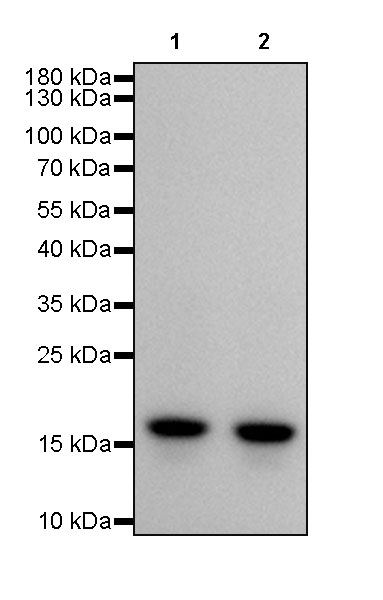
WB result of Histone H3 Rabbit mAb Primary antibody: Histone H3 Rabbit mAb at 1/1000 dilution Lane 1: HEK293 whole cell lysate 20ug Lane 2: HeLa whole cell lysate 20ug Secondary antibody: #JP20040 at 1/10000 dilution Predicted MW: 15kDa Observed MW: 17kDa Exposure time: 3s
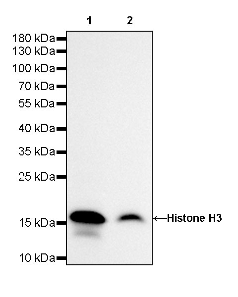
WB result of Histone H3 Rabbit mAb Primary antibody: Histone H3 Rabbit mAb at 1/1000 dilution Lane 1: NIH/3T3 whole cell lysate 20ug Lane 2: mouse brain lysate 20ug Secondary antibody: #JP20040 at 1/10000 dilution Predicted MW: 15kDa Observed MW: 17kDa Exposure time: 20s
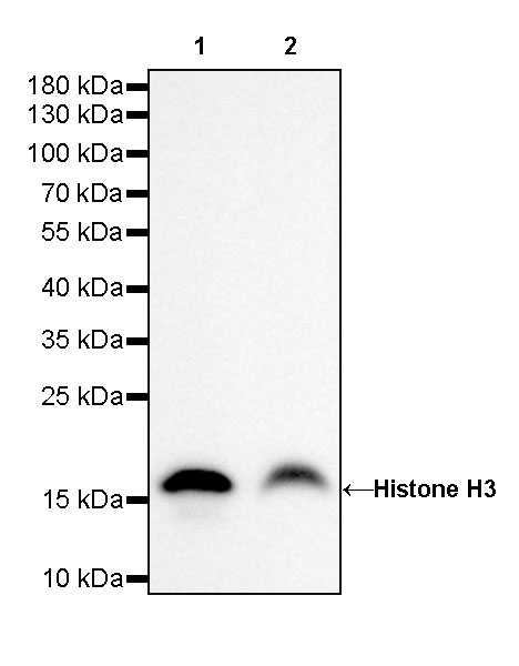
WB result of Histone H3 Rabbit mAb Primary antibody: Histone H3 Rabbit mAb at 1/1000 dilution Lane 1: C6 whole cell lysate 20ug Lane 2: rat liver lysate 20ug Secondary antibody: #JP20040 at 1/10000 dilution Predicted MW: 15kDa Observed MW: 17kDa Exposure time: 20s
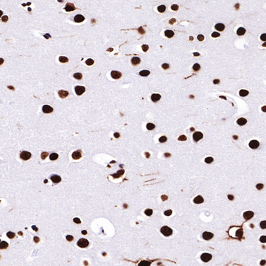
IHC shows positive staining in paraffin-embedded human cerebral cortex. Anti-Histone H3 antibody was used at 1/4000 dilution, followed by a HRP Polymer for Mouse & Rabbit IgG (ready to use). Counterstained with hematoxylin. Heat mediated antigen retrieval with Tris/EDTA buffer pH9.0 was performed before commencing with IHC staining protocol.

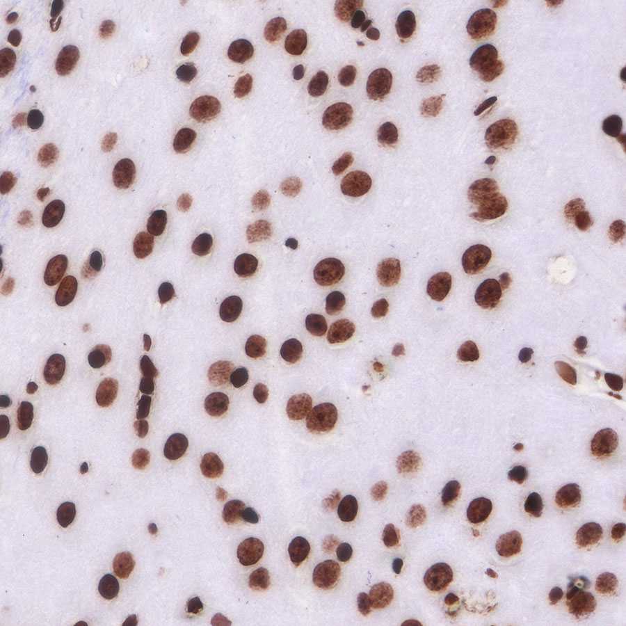
IHC shows positive staining in paraffin-embedded human spleen. Anti-Histone H3 antibody was used at 1/4000 dilution, followed by a HRP Polymer for Mouse & Rabbit IgG (ready to use). Counterstained with hematoxylin. Heat mediated antigen retrieval with Tris/EDTA buffer pH9.0 was performed before commencing with IHC staining protocol.
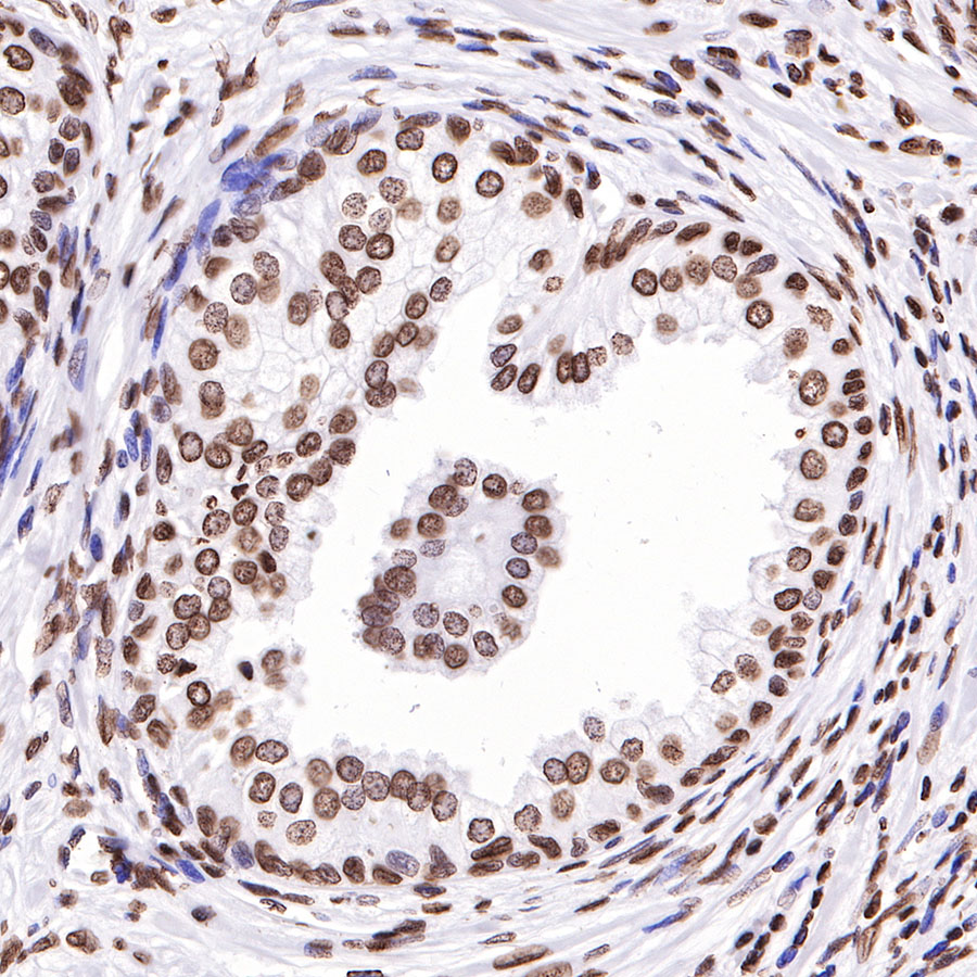
IHC shows positive staining in paraffin-embedded human prostate. Anti-Histone H3 antibody was used at 1/4000 dilution, followed by a HRP Polymer for Mouse & Rabbit IgG (ready to use). Counterstained with hematoxylin. Heat mediated antigen retrieval with Tris/EDTA buffer pH9.0 was performed before commencing with IHC staining protocol.
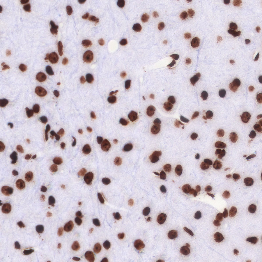
IHC shows positive staining in paraffin-embedded mouse cerebral cortex. Anti-Histone H3 antibody was used at 1/4000 dilution, followed by a HRP Polymer for Mouse & Rabbit IgG (ready to use). Counterstained with hematoxylin. Heat mediated antigen retrieval with Tris/EDTA buffer pH9.0 was performed before commencing with IHC staining protocol.
IHC shows positive staining in paraffin-embedded rat cerebral cortex. Anti-Histone H3 antibody was used at 1/4000 dilution, followed by a HRP Polymer for Mouse & Rabbit IgG (ready to use). Counterstained with hematoxylin. Heat mediated antigen retrieval with Tris/EDTA buffer pH9.0 was performed before commencing with IHC staining protocol.

ICC shows positive staining in HeLa cells. Anti-Histone H3 antibody was used at 1/500 dilution (Green) and incubated overnight at 4°C. Secondary antibody: #JP20025 at 1/1000 dilution. The cells were fixed with 100% ice-cold methanol and permeabilized with 0.1% PBS-Triton X-100. Nuclei were counterstained with DAPI (Blue). Counterstain with tubulin (red).
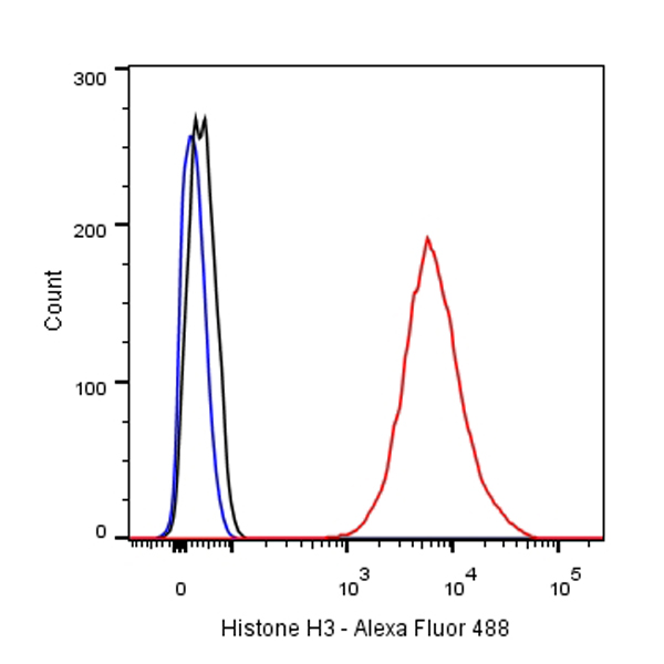
Flow cytometric analysis of HeLa cells labelling Histone H3 antibody at 1/500 (0.1ug) dilution/ (red) compared with a Rabbit monoclonal IgG (Black) isotype control and an unlabelled control (cells without incubation with primary antibody and secondary antibody) (Blue). Secondary antibody: #JP20025.