StressMarq——Alpha 突触核蛋白产品推荐(2)

StressMarq——Alpha 突触核蛋白产品推荐(2)

StressMarq——Alpha 突触核蛋白产品推荐(2)


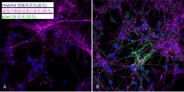
1型 PFFs 诱发iPSC-衍生神经元中的pSer129 病理现象

超声处理的 1 型 α-突触核蛋白原纤维 (目录号 SPR-322) 诱导 iPSC 衍生神经元中α-突触核蛋白丝氨酸 129 的磷酸化,这是在 PD 中观察到的典型修饰。

孵育7天后进行p-Ser129染色,1型原纤维 (目录号 SPR-322) 7天内诱发iPSC-衍生神经元中磷酸化丝氨酸129 病理现象:

StressMarq——Alpha 突触核蛋白产品推荐(2)

A) 阴性对照孔(未添加原纤维). B) 加入目录号 SPR-322 原纤维的孔

来源: Cellstate Biosciences

1型 PFFs 诱发大鼠多巴胺能神经元中的蛋白聚集和毒性

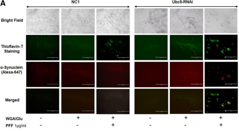
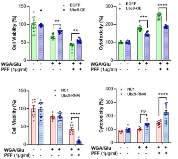
N27 大鼠多巴胺能神经元用 1 型小鼠 PFF(目录号 SPR-324)以 1 µg/ml 处理。Ubc9 过表达诱导高水平的类泛素化修饰

StressMarq——Alpha 突触核蛋白产品推荐(2)

StressMarq——Alpha 突触核蛋白产品推荐(2)

PFF 产生 α-突触核蛋白聚集(由硫磺素 T 测定)并降低细胞活力(MTT 测定)、诱导细胞毒性(LDH 测定)并增加 ROS 水平(CellROX)

研究学者使用StressMarq的小鼠 PFFs (目录号 SPR-324) 显示 α-突触核蛋白类泛素化修饰可能有神经保护功能

来源: Verma et al., https://doi.org/10.1523/ENEURO.0134-20.2020  


1型小鼠PFFs 诱发原代多巴胺能神经元中的Alpha突触核蛋白聚集

StressMarq——Alpha 突触核蛋白产品推荐(2)

研究人员使用 StressMarq 的小鼠 PFF(目录号 SPR-324)来证明特定的 CBP/p300 抑制剂可以减弱alpha 突触核蛋白聚集

来源:Hlushchuk et al. https://pubs.acs.org/doi/abs/10.1021/acschemneuro.1c00215

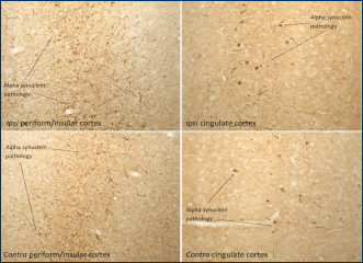
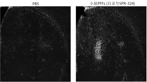
1型 PFFs 诱发大鼠脑中的 pSer129 病理现象

StressMarq 的小鼠 α 突触核蛋白 PFF(目录号 SPR-324)可在30天内诱发从注射部位扩散的病理学

StressMarq——Alpha 突触核蛋白产品推荐(2)

将 16 µg 小鼠 α-syn 1 型原纤维(目录号 SPR-324)注射到 Sprague-Dawley 大鼠纹状体一侧的两个位点。30 天后,提取大脑并进行 pSer129 固定和染色。在皮层的同侧(顶部)和对侧(底部)部分可以看到 PD 样病理的扩散。

注: performed by Atuka Inc., Toronto

StressMarq——Alpha 突触核蛋白产品推荐(2)

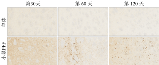
PFFs(目录号 SPR-324)两位点双边纹状体内注射至大鼠大脑,图像显示纹状体

来源: performed by Atuka Inc., Toronto

StressMarq——Alpha 突触核蛋白产品推荐(2)

将 PFF(目录号 SPR-324)通过两个位点双边纹状体注射至大鼠大脑,图像显示扣带皮层

来源: performed by Atuka Inc., Toronto


StressMarq——Alpha 突触核蛋白产品推荐(2)

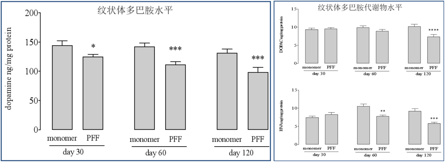
将PFF(目录号 SPR-324)从两位点双边纹状体内注射大鼠大脑后,纹状体中多巴胺和多巴胺代谢物的水平

来源: performed by Atuka Inc., Toronto

1型 PFFs 诱发小鼠脑中的 pSer129 病理现象
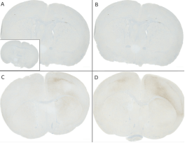
8 周大的C57/BL6 小鼠注射了超声处理的重组小鼠alpha突触核蛋白单体(目录号 SPR-323)或PFFs (目录号 SPR-324)。小鼠背侧纹状体单侧注射 (bregma AP + 0.2 mm,L +/- 2.0 mm, V – 3.0 mm) 并于注射30天后取脑切片。注射了PFF 的小鼠显示在注射位点对侧纹状体和皮质都有典型的PD pSer129 病理现象。
StressMarq——Alpha 突触核蛋白产品推荐(2) StressMarq——Alpha 突触核蛋白产品推荐(2)
(A) 1.25 uL 小鼠alpha 突触核蛋白单体 (SPR-323). (B) 2.5 uL 小鼠alpha 突触核蛋白单体 (SPR-323). (C) 2.5 ug alpha 突触核蛋白 PFFs (SPR-324). (D) 5 ug alpha 突触核蛋白 PFFs (SPR-324). 内嵌图: PBS (阴性对照)

(A) 对侧皮质. (B) 同侧皮质.

(C) 对侧纹状体. (D) 同侧纹状体. 

来源: Porsolt, France

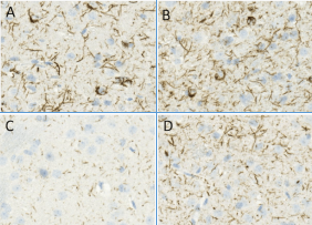
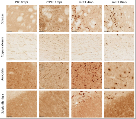
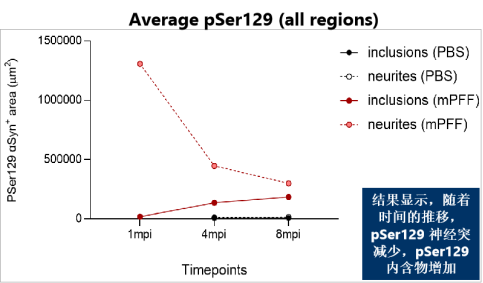
1型 PFFs 诱发C57BL/6小鼠中的 pSer129 病理学

8-9 周大的C57BL/6 小鼠注射了 4 µg 超声处理的重组小鼠 alpha 突触核蛋白原纤维 (目录号 SPR-324)。

免疫组织学分析显示在小鼠大脑的不同区域检测到 pSer129 α 突触核蛋白病理学。

StressMarq——Alpha 突触核蛋白产品推荐(2)

StressMarq——Alpha 突触核蛋白产品推荐(2)

来源: KU Leuven, Belgium

1型 PFFs 诱发57BL/6 小鼠中的神经炎症

StressMarq——Alpha 突触核蛋白产品推荐(2)

注射后4个月时检测 C57BL/6 小鼠大脑注射纹状体中的 GFAP+ 星形胶质细胞

来源: KU Leuven, Belgium

1型 PFF被SH-SY5Y 细胞吸收后在14天内转移到了神经元iPSC中
StressMarq——Alpha 突触核蛋白产品推荐(2)
Alpha 突触核蛋白 PFF 信号(目录号 SPR-322,红色)与 iPSC(洋红色)信号相关,表明来自 SH-SY5Y 细胞(绿色)的 PFF 已转移。

来源: Neuronal cultures and ICC performed by Cellstate Biosciences (USA)

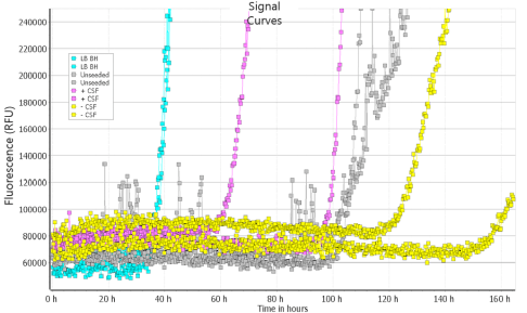
2型单体用于脑脊液聚集检测

2 型单体(目录号 SPR-316)聚集检测可区分患病样品和正常样品。

StressMarq——Alpha 突触核蛋白产品推荐(2)

来源:RT-QuIC assay by Dr. Alison Green and Graham Fairfoul (Green Lab), University of Edinburgh, UK

2型 PFF 影响 SH-SY5Y 成神经细胞瘤细胞膜

美国加州州立大学的研究人员研究了2型α突触核蛋白PFFs(目录号SPR-317)暴露对SH-SY5Y神经母细胞瘤细胞膜的影响。将不同浓度的α突触核蛋白PFFS添加到神经母细胞瘤细胞中48小时。 使用扫描离子电导显微镜对α 突触核蛋白 PFF 与细胞膜的相互作用进行表征。

● SICM 图像表明,随着原纤维浓度的增加,膜粗糙度显著增加。

● 48小时内,PFF继续在细胞膜上积累

● XTT 细胞活力和 LDH 细胞毒性测定表明 PFF 没有诱导大量细胞死亡

● Alpha 突触核蛋白 PFFs 影响细胞膜并破坏细胞功能

StressMarq——Alpha 突触核蛋白产品推荐(2)

PFF(目录号 SPR-317)处理 48 小时后 SH-SY5Y 细胞膜的代表性 SICM 图像。

来源: Feng et al. https://doi.org/10.1021/acschemneuro.2c00478


更多详情请联系Stressmarq中国代理商——上海金畔生物

原位杂交(FISH)标记及检测

原位杂交(FISH)标记及检测

本篇给大家介绍的是原位杂交(FISH)标记及检测相关试剂产品,如需获取这些产品的详细信息,可以点击产品名称或产品货号,了解产品说明书及产品信息,也可联系ENZO代理商上海金畔生物。

原位杂交(FISH)标记及检测产品列表

原位杂交(FISH)标记及检测 产品名称 货号
Nick Translation DNA Labeling System 2.0 ENZ-GEN111
SEEBRIGHT® Gold 550 dUTP ENZ-42521
SEEBRIGHT® Green 496 dUTP ENZ-42831
SEEBRIGHT® Red 650 dUTP ENZ-42522

如需了解更多详情,请联系Enzo Life Sciences国内代理商-上海金畔生物!

MITO-ID®线粒体膜电位检测试剂盒——ENZO热销产品

MITO-ID®线粒体膜电位检测试剂盒——ENZO热销产品

线粒体膜电位(Mitochondrial Membrane Potential,MMP)是判定细胞健康程度、线粒体膜通透性和细胞凋亡的一个重要指标,MMP的丧失通常与细胞凋亡的早期阶段有关。评估线粒体功能状态的基于细胞的检测方法正在成为阐明线粒体活动在药物诱导毒性、细胞凋亡级联以及其他细胞和生化过程中的作用的有用工具。

Enzo Life SciencesMITO-ID® Membrane potential detection kit包含一种双发射阳离子染料,用于检测活细胞中的线粒体膜电位(MMP)。在有能量或活跃的细胞中,MITO-ID® 膜电位试剂因其相对负电荷而在线粒体中迅速聚集成发橙色荧光的聚合体,而在细胞质中则以发绿色荧光的单体存在。然而,在MMP受损的细胞中,MITO-ID®膜电位试剂主要以绿色荧光单体存在于整个细胞质中,在线粒体中不再表现出橙色荧光。


作用机制

该染料的基本化学结构由高度共轭的部分组成,使正电荷广泛离域。该染料能够选择性地进入线粒体,当膜电位增加时,它的颜色会从绿色可逆地变为橙色(双发射电位探针)。这种光物理特性是由于在膜极化时可逆地形成J-聚集体,导致在490nm处激发时,发射光从~530nm转移到590nm。因此,具有低膜电位的线粒体将积累低浓度的染料,并表现出绿色荧光,而更高极化的线粒体将表现出橙色的荧光。


MITO-ID®线粒体膜电位检测试剂盒——ENZO热销产品

 MITO-ID® Membrane potential detection kit线粒体膜电位检测试剂盒


产品特点

● 耐光双发射染料,能够根据线粒体膜电位状态发出绿色或橙色荧光

● JC-1荧光染料灵敏10倍,并具有卓越的水溶性

● 无需洗涤步骤

● 适用于化学/环境毒性筛选

● 适用于高通量应用


实验示例

MITO-ID®线粒体膜电位检测试剂盒——ENZO热销产品

1. MITO-ID® Membrane Potential reagentHeLa细胞的线粒体进行染色,并通过荧光显微镜进行观察。橙色荧光聚集体定位于线粒体(橙色通道),而绿色荧光单体主要定位于细胞质(FITC通道)。
 

MITO-ID®线粒体膜电位检测试剂盒——ENZO热销产品

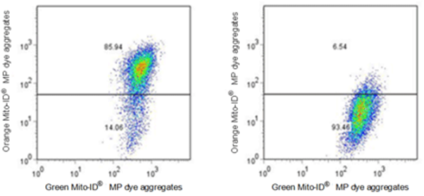
2. 对照组和实验组细胞的流式细胞仪分析。未经处理的Jurkat细胞(左)与用1μM CCCP处理15分钟的Juekat细胞(右),用MITO-ID® Membrane Potential reagent对细胞进行染色,并使用流式细胞仪检测。


产品信息

产品货号

ENZ-51018-0025/ ENZ-51018-K100

产品名称

MITO-ID® Membrane potential detection kit 

规格

1*25tests/1*100tests

短期保存

-20°C

长期保存

-80°C

试剂盒组分

MITO-ID® MP Detection Reagent

Necrosis Detection Reagent

CCCP Control

10X Assay Buffer 1

50X Assay Buffer 2

应用

Flow Cytometry, Fluorescence microscopy, Fluorescent detection, HTS


部分产品引用文献

1. Discovery of the 3-Amino-1,2,4-triazine-Based Library as Selective PDK1 Inhibitors with Therapeutic Potential in Highly Aggressive Pancreatic Ductal Adenocarcinoma: D. Carbone, et al.; Int. J. Mol. Sci. 24, 3679 (2023) 

2. Gene signature predicting recurrence in oral squamous cell carcinoma is characterized by increased oxidative phosphorylation: J.K. Noh, et al.; Mol. Oncol. 17, 134 (2023) 

3. Cu(I) and Cu(II) Complexes Based on Lonidamine-Conjugated Ligands Designed to Promote Synergistic Antitumor Effects: F.D. Bello, et al.; Inorg. Chem. 61, 4919 (2022) 

4. Mitochondrial membrane potential-enriched CHO host: a novel and powerful tool for improving biomanufacturing capability: L. Chakrabarti, et al.; MAbs 14, 2020081 (2022)

5. New glycoconjugation strategies for Ruthenium(II) arene complexes via phosphane ligands and assessment of their antiproliferative activity: D. Iacopini, et al.; Bioorg. Chem. 126, 105901 (2022)

6. New tricyclic systems as photosensitizers towards triple negative breast cancer cell: M. Barreca, et al.; Arch. Pharm. Res. 45, 806 (2022) 

7. Role of caspase-8 and/or-9 as biomarkers that can distinguish the potential to cause toxic-and immune related-adverse event, for the progress of acetaminophen-induced liver injury: T. Noda, et al.; Life Sci. 294, 120351 (2022), Application(s): Microplate reader 

8. Unveiling the Potential of Innovative Gold(I) and Silver(I) Selenourea Complexes as Anticancer Agents Targeting TrxR and Cellular Redox Homeostasis: M.D. Franco, et al.; Chemistry 28, e202201898 (2022) 

9. Altered inflammatory response in FMRP-deficient microglia: J.M. Parrott, et al.; iScience 24, 103293 (2021), Application(s): Fluorescence microscopy 

10. ApoE4 impairs neuron-astrocyte coupling of fatty acid metabolism: G. Qi, et al.; Cell. Rep. 34, 108572 (2021), Application(s): Microplate reader 

11. Botrytis cinerea methyl isocitrate lyase mediates oxidative stress tolerance and programmed cell death by modulating cellular succinate levels: L. Oren-Young, et al.; Fungal Genet. Biol. 146, 103484 (2021), Application(s): Conidia (fungus spore); microscopy 

12. Copper (II) complexes containing natural flavonoid pomiferin show considerable in vitro cytotoxicity and anti-inflammatory effects: J. Vanco, et al.; Int. J. Mol. Sci. 22, 7626 (2021), Application(s): Flow cytometry

13. Depletion of mitochondrial components from extracellular vesicles secreted from astrocytes in a mouse model of fragile X syndrome: B.G. Ha, et al.; Int. J. Mol. Sci. 22, 410 (2021), Application(s): Fluorescence microscopy

14. Inhibiting autophagy targets human leukemic stem cells and hypoxic AML blasts by disrupting mitochondrial homeostasis: K.M. Dykstra, et al.; Blood Adv. 5, 2087 (2021)

15. Selective striatal cell loss is ameliorated by regulated autophagy of the cortex: K. Cho & G.W. Kim; Life Sci. 282, 119822 (2021), Application(s): Flow cytometry


MITO-ID® Membrane potential cytotoxicity kit线粒体膜电位细胞毒性检测试剂盒

Enzo Life SciencesMITO-ID® Membrane potential cytotoxicity kit利用阳离子双发射染料检测线粒体膜电位(MMP)的波动,该染料在细胞质中以绿色荧光单体的形式存在,在线粒体中以橙色荧光J聚集体的形式聚集。具有低膜电位的线粒体将积累低浓度的染料并呈现绿色荧光,而更高度极化的线粒体将呈现橙红色荧光。随着线粒体功能受损加剧,细胞表现出从橙色荧光到绿色荧光的转变。该试剂盒是一种独特的HTS测定法,无需洗涤或去除培养基即可实时监测线粒体膜电位。


产品特点

● 灵敏度是JC-1荧光染料的10倍,并具有卓越的水溶性

● 具备光稳定性的双发射染料

● 无需漂洗/换液步骤

● 单独的MITO-ID®检测可用于检测线粒体质量

● 可在较低的药物/剂量浓度下检测细胞毒性

● 没有使用JC-1染料时出现的溶剂伪影

● 适用于高通量应用


实验示例
MITO-ID®线粒体膜电位检测试剂盒——ENZO热销产品

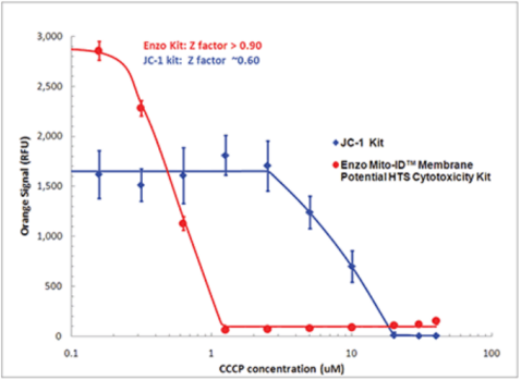
1. 检测线粒体紊乱的灵敏度是JC-110倍。使用MITO-ID®膜电位染料(红色)或JC-1(蓝色)在经CCCP处理的HeLa细胞中评估线粒体膜电位(MMP)。使用传统的荧光酶标仪检测,结果显示MMP 随着CCCP浓度的增加而减少,橙色荧光减少。染料的水溶性优化和无需洗涤的实验方案最大限度减小了可变性,因此Z因子(>0.9)高于使用JC-1.
 

MITO-ID®线粒体膜电位检测试剂盒——ENZO热销产品

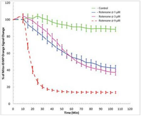
2. 在药物筛选中实时检测有丝分裂毒性。使用BioTek Synergy™ Mx荧光酶标仪对线粒体膜电位变化的时间进程研究。HeLa细胞与MITO-ID®膜电位染料在室温下孵育30分钟(不去除血清或培养基)。添加鱼藤酮分别达到1µM3µM9µM的浓度。橙色信号的减少证明染料对鱼藤酮有反应。


产品信息

产品货号

ENZ-51019-KP002

产品名称

MITO-ID® Membrane potential cytotoxicity kit

规格

1 Kit

短期保存

-20°C

长期保存

-80°C

试剂盒组分

MITO-ID® MP Detection Reagent, 200 μL

CCCP Control, 100 μL

10X Assay Buffer 1: 2.5 mL

50X Assay Buffer 2: 0.5 mL

应用

HTS
Microplate


部分产品引用文献

1. Novel Silver Complexes Based on Phosphanes and Ester Derivatives of Bis(pyrazol-1-yl)acetate Ligands Targeting TrxR: New Promising Chemotherapeutic Tools Relevant to SCLC Management: M. Pellei, et al.; Int. J. Mol. Sci. 24, 4091 (2023) 

2. FUNDC1 regulates receptor-mediated mitophagy independently of the PINK1/Parkin-dependent pathway in rotenone-treated SH-SY5Y cells: S.Y. Park, et al.; Food Chem. Toxicol. 137, 111163 (2020), Application(s): Fluorescence microscopy and microplate reader

3. Inhibitory role of TRIP-Br1/XIAP in necroptosis under nutrient/serum starvation: Z. Sandag, et al.; Mol. Cells 43, 236 (2020) 

4. NAD hydrolysis by the tuberculosis necrotizing toxin induces lethal oxidative stress in macrophages: D. Pajuelo, et al.; Cell. Microbiol. 22, e13115 (2020), Application(s): THP-1 macrophages; microplate reader

5. Impaired autophagic and mitochondrial functions are partially restored by ERT in Gaucher and Fabry diseases: M.M. Ivanova, et al.; PLoS One 14, e0210617 (2019), Application(s): Fluorescence microscopy 

6. Inhibitory role of AMPactivated protein kinase in necroptosis of HCT116 colon cancer cells with p53 null mutation under nutrient starvation: D.T. Le, et al.; Int. J. Oncol. 54, 702 (2019) 

7. ROS as a novel indicator to predict anticancer drug efficacy: T. Zaidieh, et al.; BMC Cancer 19, 1224 (2019) 

8. TREM1/3 deficiency impairs tissue repair after acute kidney injury and mitochondrial metabolic flexibility in tubular epithelial cells: A. Tammaro, et al.; Front. Immunol. 10, 1469 (2019), Application(s): Microplate reader

9. Anti-cancerous effect of cis-khellactone from Angelica amurensis through the induction of three programmed cell deaths: S. Jung, et al.; Oncotarget 9, 16744 (2018), Application(s): Microplate reader 

10. Blue light phototoxicity toward human corneal and conjunctival epithelial cells in basal and hyperosmolar conditions: V. Marek, et al.; Free Radic. Biol. Med. 126, 27 (2018), Application(s): Microplate reader

11. cGAS drives noncanonical-inflammasome activation in age-related macular degeneration: N. Kerur, et al.; Nat. Med. 24, 50 (2018)

12. Cytotoxicity of propofol in human induced pluripotent stem cell-derived cardiomyocytes: K. Kido, et al.; J. Anesth. 32, 120 (2018), Application(s): Microplate reader

13. Development of novel amino-quinoline-5, 8-dione derivatives as NAD (P) H: quinone oxidoreductase 1 (NQO1) inhibitors with potent antiproliferative activities: Y. Ling, et al.; Eur. J. Med. Chem. 154, 199 (2018)

14. Inhibition of apoptosis using exosomes in Chinese hamster ovary cell culture: S. Han, et al.; Biotechnol. Bioeng. 115, 1331 (2018)

15. Light action spectrum on oxidative stress and mitochondrial damage in A2E-loaded retinal pigment epithelium cells: M. Marie, et al.; Cell Death Disc. 9, 287 (2018)


详情请咨询Enzo Life Sciences代理——上海金畔生物
StressMarq——Alpha 突触核蛋白产品推荐(1)

StressMarq——Alpha 突触核蛋白产品推荐(1)

 StressMarq——Alpha 突触核蛋白产品推荐(1)

前体原纤维 (PFFs) 来源物种 表达系统 突变型/WT 目录号
Alpha 突触核蛋白PFF (1) E. Coli WT SPR-322
Alpha 突触核蛋白PFF (1) (ATTO 594 标记的) E. Coli WT SPR-322-A594
Alpha 突触核蛋白PFF (1) 小鼠 E. Coli WT SPR-324
Alpha 突触核蛋白PFF (2) E. Coli WT SPR-317
Alpha 突触核蛋白N-末端乙酰化的PFF (1) E. Coli WT SPR-332
Alpha 突触核蛋白A53T 突变型 PFF (1) E. Coli A53T SPR-326
Alpha 突触核蛋白S87N 突变型 PFF E. Coli S87N SPR-500
Alpha 突触核蛋白PFF 大鼠 E. Coli WT SPR-482
Alpha 突触核蛋白纤丝 (未成熟的原纤维)* E. Coli WT SPR-450
Tau-352 (胚胎 0N3R)  Alpha 突触核蛋白共聚集体原纤维 E. Coli WT SPR-494
Tau-441 (2N4R)  Alpha 突触核蛋白共聚集体原纤维 E. Coli WT SPR-495
Alpha Synuclein S129A 突变型PFF E. Coli S129A SPR-506
Alpha Synuclein TNG (A53T, S87N, N103G) 突变型PFF E. Coli A53T, S87N, N103G SPR-504

* 由于操作不同可能含有少量的不溶纤维。


寡聚体 来源物种 表达系统 突变型/WT 目录号
动态学稳定的 Alpha 突触核蛋白寡聚体 E. coli WT SPR-484
盐酸多巴胺稳定的 Alpha 突触核蛋白寡聚体 E. coli WT SPR-466
EGCG稳定的 Alpha 突触核蛋白寡聚体 E. coli WT SPR-469
单体 来源物种 表达系统 突变型/WT 目录号
Alpha 突触核蛋白单体 (1) E. coli WT SPR-321
Alpha 突触核蛋白单体 (2) E. coli WT SPR-316
Alpha 突触核蛋白单体 (1) 小鼠 E. coli WT SPR-323
Alpha 突触核蛋白A53T 突变型单体 (1) E. coli A53T SPR-325
Alpha 突触核蛋白S87N 突变型单体 E. coli S87N SPR-499
Alpha 突触核蛋白N-末端乙酰化的单体 (1) E. coli WT SPR-331
Alpha 突触核蛋白单体 大鼠 E. coli WT SPR-481
Alpha Synuclein S129A 突变型单体 E. coli S129A SPR-505
Alpha Synuclein TNG (A53T, S87N, N103G) 突变型单体 E. coli A53T, S87N, N103G SPR-503

Alpha 突触核蛋白产品类型:PFF

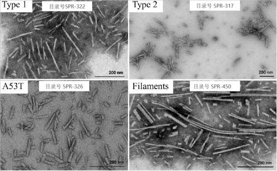
不同类型蛋白看起来形态相似, 但是特性不同

StressMarq——Alpha 突触核蛋白产品推荐(1)

来源: 

1型原纤维(左上) 和纤丝(右下) 的EM来自加拿大维多利亚大学。

2型原纤维 (右上) 和A53T 突变型原纤维 (左下) 的EM 来自加拿大英属哥伦比亚大学。


Alpha 突触核蛋白产品类型: PFFs

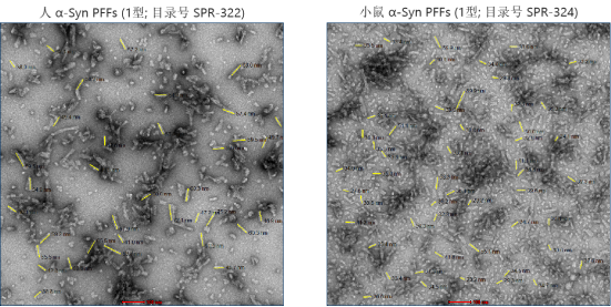
StressMarq提供人、小鼠和大鼠Alpha 突触核蛋白PFFs

StressMarq——Alpha 突触核蛋白产品推荐(1)

原纤维在浴式超声仪 (QSonica) 中以 80% 振幅进行超声处理(开启 30 秒,关闭 30 秒,10°C 下 30 个循环)。原纤维用乙酸双氧铀进行负染色,并在加拿大多伦多大学的 Talos L120C 透射电子显微镜中观察。

来源:Atuka Inc.。


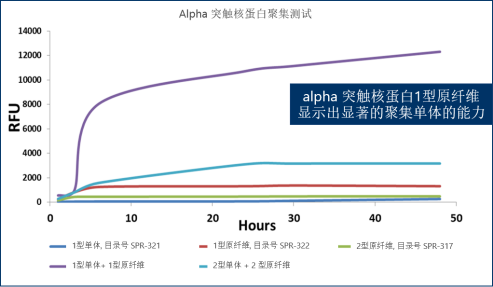
硫磺素 T 聚集测试

StressMarq——Alpha 突触核蛋白产品推荐(1)

单体浓度: 100 µM;原纤维浓度: 10 µM

来源:StressMarq Biosciences 内部数据

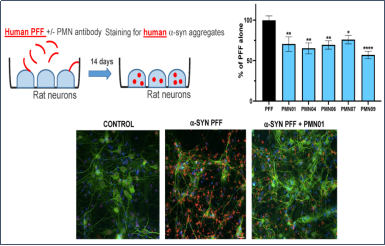
单克隆抗体选择性靶向病理性人 Alpha 突触核蛋白PFFs

不列颠哥伦比亚大学的研究人员使用 StressMarq 的 Alpha 突触核蛋白原纤维(目录号 SPR-322)通过计算设计了一种小环肽,该肽表现出与原纤维相同的神经毒性和聚集活性。他们通过肽支架免疫产生了有高度选择性的单克隆抗体,能够抑制α突触核蛋白原纤维的致病作用。这项研究可能在α突触核蛋白靶向治疗突触核蛋白病方面具有重要应用。


StressMarq——Alpha 突触核蛋白产品推荐(1)

mAb 抑制 PFF 摄取和细胞内聚集体形成。将原代大鼠海马神经元培养物暴露于超声处理的人 PFF(目录号 SPR-322,1 μg/mL)中,不含或含 mAb(0.05 μM,PMN09 是0.25 μM)。 14天后染色处理,神经元标记物MAP2 (绿色), 人 α-syn 聚集体(红色)和细胞核(蓝色)染色。


StressMarq——Alpha 突触核蛋白产品推荐(1)

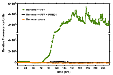
mAb 抑制 PFF 聚集活性。在单独添加 α-syn 单体 (100 mM) 后或在超声处理的人 PFF (10 nM) 作为引种剂的情况下,不添加或添加 mAb (0.1 nM),每小时用 ThT 荧光测定(每次读数前摇动 30 秒)跟踪聚集情况。

来源:Gibbs, E. et al. https://doi.org/10.3390/biomedicines10092168

帕金森样病理学在初级神经元中的诱发

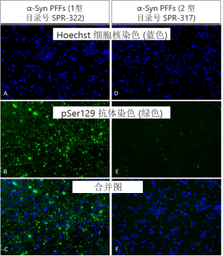
超声处理的 1 型 α-突触核蛋白原纤维 (4 µg/ml) 诱导原代大鼠神经元中丝氨酸 129 的磷酸化,这是在 PD 中观察到的典型修饰现象。

孵育 14 天后进行 p-Ser129 染色:

◆ 1型原纤维在14天内诱发原代大鼠神经元中的帕金森样病理学

◆ 2型原纤维没有这个现象

StressMarq——Alpha 突触核蛋白产品推荐(1)

来源: Associate Professor Rehana Leak, Duquesne University, USA


更多详情请联系Stressmarq中国代理商——上海金畔生物
Cayman chemical公司免疫与炎症研究试剂

Cayman chemical公司免疫与炎症研究试剂

Cayman chemical公司成立于1980年,总部位于美国密歇根州。该公司汇集了众多生化合成领域的专家,为各研究领域的科学家们提供各种生化研究工具,如为癌症,氧化损伤,表观遗传学,神经科学,炎症,代谢等许多研究领域提供检测试剂盒,用于检测类花生酸、自由基生物标志物、环核苷酸、细胞因子、激素和一氧化氮,以及各种高质量的生化制品,包括类花生酸、一氧化氮试剂、小分子抑制剂以及各种相关的脂类、脂肪酸、酶和抗体。

金畔作为国内代理商,已代理Cayman品牌15余年,为国内众多科研院校及工业客户提供了长期的优质服务。本资料为综合筛选的Cayman热销品类展示,如您需要更多资讯,请联系我们!

免疫与炎症研究产品

产品名称 货号 说明
Resolvin D1 ELISA Kit 500380 脂类炎症介质检测
Maresin 1 ELISA Kit 501150 脂类炎症介质检测
Leukotriene B4 EIA Kit 520111 脂类炎症介质检测
Lipoxin A4 ELISA Kit 590410 脂类炎症介质检测

Prostaglandin 

E2 EIA Kit – Monoclonal

514010 脂类炎症介质检测

Prostaglandin E2 

Express EIA Kit

500141 脂类炎症介质检测

Anti-Ovalbumin IgE 

(mouse) EIA Kit

500840 过敏
BAY-u3405 10156 过敏
Histamine EIA Kit 589651 过敏
产品名称 货号 说明
Phagocytosis Assay Kit 

(IgG FITC)

500290 天然免疫-吞噬作用
2’3′-cGAMP ELISA Kit 501700 天然免疫-STING
Cyclic di-GMP ELISA Kit 501780 天然免疫-STING
Cyclic di-AMP ELISAKit 501960  天然免疫-STING
COX-2 (human 

recombinant)

60122 环氧合酶活性及

抑制剂检测

COX(ovine/human) Inhibitor

Screening Assay Kit

560131

环氧合酶活性及

抑制剂检测

COX-1 (human 

recombinant)

17616

环氧合酶活性及

抑制剂检测

更多Cayman产品详情,请咨询代理商-上海金畔生物

Enzo Life Sciences表观遗传学热销产品列表

Enzo Life Sciences表观遗传学热销产品列表

Enzo Life Sciences成立于1976年,总部设于美国纽约,在研发、生产、销售生物研究试剂方面拥有40多年的成功经验,涉及靶标鉴定及验证、高通量分析、基因表达分析、蛋白质检测及细胞分析等相关领域,可提供蛋白、抗体、多肽、小分子、标签探针染料及试剂盒等一万余种产品,本篇仅介绍部分热销产品,上海金畔生物代理Enzo Life Sciences 15余年,为您提供专业的售前售后服务,如需咨询更多详情,欢迎联系上海金畔生物!

本篇是对于Enzo Life Sciences表观遗传学热销产品进行展示,如需了解该产品的详细信息,可以在本站搜索框搜索产品的名称或产品的货号,即可查看该产品的详细信息。

产品名称 货号
样本制备 EPIXTRACT® Total Histone Extraction Kit ENZ-45013/45014
EPIXTRACT® Nuclear Protein Isolation Kit ENZ-45015/45016
EPIXTRACT® DNA Isolation Kit for Plasma/Serum ENZ-45018
样本转换(甲基化) Express DNA Methylation Kit ENZ-45001
Blood & Tissue DNA Methylation Kit ENZ-45004
BIOARRAY™ 5-hmC Methylation Kit ENZ-45011
脱乙酰作用 FLUOR DE LYS® HDAC fluorometric activity assay kit BML-AK500
FLUOR DE LYS® HDAC Cellular Activity Assay Kit BML-AK503
FLUOR DE LYS® SIRT3 fluorometric drug discovery assay kit BML-AK557
FLUOR DE LYS® HDAC6 fluorometric drug discovery kit BML-AK516
泛素化及类泛素化试剂盒及蛋白 SUMOylation Kit BML-UW8955
SUMO-QAPTURE-T® Kit BML-UW1000A
UBI-QAPTURE-Q® Kit BML-UW8995A
Auto-Ubiquitinylation Kit BML-UW0970
NEDDylation Kit BML-UW0590
Ubiquitin Activating Kit BML-UW0400A
Ubiquitin Conjugating Kit (HeLa lysate-based)  BML-UW9915
Ubiquitinylation Kit BML-UW9920
Ubiquitin aldehyde (recombinant) BML-UW8450
Ubiquitin activating enzyme E1, (recombinant) (His-tag) BML-UW9410
Mg2+/ATPActivating Solution BML-EW9805
Mono- and polyubiquitinylated conjugates recombinant monoclonal antibody ENZ-ABS840
甲基化及羟甲基化检测试剂盒 5-Methylcytosine DNA ELISA Kit ADI-900-224
5-Hydroxymethylcytosine DNA ELISA Kit ADI-900-225

如需了解更多详情,请联系Enzo Life Sciences国内代理商-上海金畔生物!

EpiCypher新品推荐——SNAP-CUTANA™ DYKDDDDK Tag Panel

EpiCypher新品推荐——SNAP-CUTANA™ DYKDDDDK Tag Panel

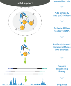
SNAP-CUTANA™ DYKDDDDK Tag Panel是 CUT&RUN 的加标对照,提供了一种用于验证抗 FLAG®* 抗体的分析内对照,并证实了涉及FLAG表位标记的染色质蛋白的CUT&RUN反应的成功。这种重要的阳性对照可指导故障排除,以区分 FLAG 表位标记问题(包括转基因表达、标记蛋白的染色质结合、标签的溶剂可及性等)与 CUT&RUN 工作流程中的技术故障。该panel由两个包含未修饰组蛋白H3或3xDYKDDDDK-H3融合的核小体组成,每个核小体都包裹有两个独特的条形码DNA模板(A和B,用于内部技术复制)。核小体单独与顺磁珠偶联并汇集到单个panel中,方便一步加标到CUT&RUN反应。在添加抗 DYKDDDDK 或 IgG 阴性对照抗体之前,将 panel 与 ConA 固定的细胞一起添加(参见应用说明和表 1)。pAG-MNase释放基因组染色质和条形码核小体取决于所使用抗体的特异性。测序后,回收的 DYKDDDDK 与未修饰核小体的相对读取计数提供了靶向与脱靶回收率的定量指标(图 2),从而衡量实验成功率并指导故障排除工作。有关工作流集成、预期结果、数据分析和故障排除的详细信息,请参阅最新的CUTANA™ CUT&RUN方法(https://www.epicypher.com/resources/protocols/cutana-cut-and-run-kit-manual)和SNAP-CUTANA™ Spike-in(https://www.epicypher.com/resources/protocols/)用户指南。

*FLAG® is a registered trademark of Merck KGaA, Darmstadt, Germany and ANTI-FLAG is a trademark of Sigma-Aldrich Co. LLC.


保存温度

自收到之日起,-20℃下可稳定储存6个月。较低的温度会导致冻结,并会永久损坏磁珠。


验证数据

EpiCypher新品推荐——SNAP-CUTANA™ DYKDDDDK Tag Panel

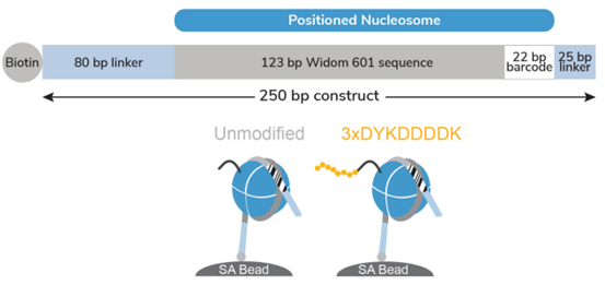
Figure 1: Schematic of SNAP-CUTANA™ DYKDDDDK Tag Panel

The DYKDDDDK Tag Panel contains two nucleosomes – one has an H3 tail fusion to a 3xDYKDDDDK Tag epitope and one is an unmodified control. Both octamers are wrapped with two uniquely barcoded DNA templates (A and B). Each 250 bp DNA template contains a 123 bp 601 nucleosome positioning sequence (gray) [1], a unique 22 bp DNA-barcode (white; 4 barcodes total), and a 5’ biotin-TEG. The 5’ and 3’ linkers (blue) are compatible with cleavage by pAG-MNase (EpiCypher 14-104815-1016) during CUT&RUN. The nucleosomes are individually pre-conjugated to paramagnetic beads and pooled for convenient use.

EpiCypher新品推荐——SNAP-CUTANA™ DYKDDDDK Tag Panel

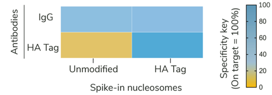
Figure 2: SNAP-CUTANA™ DYKDDDDK Tag Panel provides an in-assay control for CUT&RUN 

reactions targeting FLAG-tagged proteins

CUT&RUN was performed as described in Figure 5. CUT&RUN sequencing reads were aligned to the unique DNA barcodes corresponding to each nucleosome in the SNAP-CUTANA™ DYKDDDDK Tag Panel. Data are expressed as a percent relative to on-target recovery (DYKDDDDK Tag set to 100%) or total counts (IgG). IgG antibody results demonstrate equal loading of unmodified and epitope nucleosomes in the panel. DYKDDDDK Tag antibody results show selective enrichment of the DYKDDDDK Tag spike-in nucleosomes, validating all CUT&RUN steps, including DYKDDDDK antibody binding, pAG-MNase cleavage, and wash conditions.

EpiCypher新品推荐——SNAP-CUTANA™ DYKDDDDK Tag Panel

Table 1: Recommended SNAP-CUTANA™ DYKDDDDK Tag Panel Spike-in dilution for CUT&RUN reactions of varying starting cell number.

EpiCypher新品推荐——SNAP-CUTANA™ DYKDDDDK Tag Panel

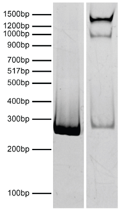
Figure 3: DNA gel data

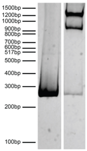
Nucleosomes in the SNAP-CUTANA™ DYKDDDDK Tag Panel were resolved via native PAGE and stained with ethidium bromide to confirm intact nucleosome assembly. Lane 1: Free 250 bp DNA used in nucleosome assembly (100 ng). Lane 2: Intact nucleosomes (400 ng).

EpiCypher新品推荐——SNAP-CUTANA™ DYKDDDDK Tag Panel

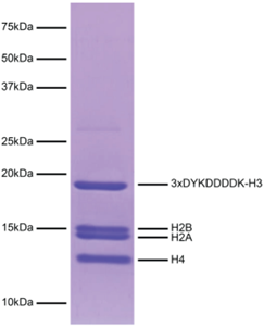
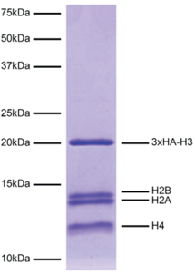
Figure 4: Protein gel data

Coomassie stained SDS-PAGE gel of the nucleosome containing a 3xDYKDDDDK-H3 fusion (1 μg) in the SNAP-CUTANA DYKDDDDK Tag Panel demonstrates the purity of histones in the preparation. Sizes of molecular weight markers and positions of the core histones (H2A, H2B, 3xDYKDDDDK-H3, and H4) are indicated.

EpiCypher新品推荐——SNAP-CUTANA™ DYKDDDDK Tag Panel

Figure 5: CUT&RUN methods

CUT&RUN was performed on 500k MDA-MB-231 native cells stably expressing 3xFLAG-tagged GATA3 [1]** using the CUTANA™ ChIC/CUT&RUN Kit v3 (EpiCypher 14-1048). SNAP-CUTANA™ DYKDDDDK Tag Panel was added just prior to the addition of either DYKDDDDK Tag (0.05 μg; EpiCypher 13-2031) or IgG negative control (0.5 μg; EpiCypher 13-0042) antibodies. Library preparation was performed with 5 ng of DNA (or the total amount recovered if less than 5 ng) using the CUTANA™ CUT&RUN Library Prep Kit (EpiCypher 14-1001/14-1002). Libraries were run on an Illumina NextSeq2000 with paired-end sequencing (2×50 bp). Data were aligned to the hg19 genome using Bowtie2. Data were filtered to remove duplicates, multi-aligned reads, and ENCODE DAC Exclusion List regions.

**Thanks to Dr. Takaku (UND) for 3xFLAG-GATA3-3xHA MDA-MB-231 cells.

订购详情

货号

产品名称

规格

19-5001

SNAP-CUTANA™ DYKDDDDK Tag Panel

50 Reactions


参考文献

[1] Lowary & Widom J. Mol. Biol. (1998). PMID: 9514715

[2] Takaku et al. Genome Biol. (2016). PMID: 26922637


如需了解更多详细信息或相关产品,

请联系EpiCypher中国代理商-上海金畔生物 

EpiCypher新品推荐——SNAP-CUTANA™ HA Tag Panel

EpiCypher新品推荐——SNAP-CUTANA™ HA Tag Panel

SNAP-CUTANA™HA Tag Panel提供了用于验证抗HA抗体和确认涉及HA表位标记的染色质蛋白的成功CUT&RUN反应的检测控制。这一重要的阳性对照指导排除故障,以区分HA表位标签问题(包括转基因表达、标记蛋白的染色质结合、标签的溶剂可及性等)与CUT&RUN工作流程中的技术故障。该面板由两个包含未修饰组蛋白H3或3xHA-H3融合的核小体组成,每个核小体都包裹有两个独特的条形码DNA模板(A和B,用于内部技术复制)。核小体分别与顺磁珠偶联,并汇集成一个Panel,方便一步插入到切割和运行反应。在添加抗ha或IgG阴性对照抗体之前,将该检测组合与ConA固定的细胞一起添加(见应用说明和表1)。pAG-MNase释放基因组染色质和条形码核小体取决于所用抗体的特异性。测序后,恢复的HA与未修饰的核小体的相对读长计数提供了在靶恢复与脱靶恢复的定量指标(图2),从而衡量实验的成功率,并指导故障排除工作。有关工作流集成、预期结果、数据分析和故障排除的详细信息,请参阅最新的CUTANA™ CUT&RUN方法(https://www.epicypher.com/resources/protocols/cutana-cut-and-run-kit-manual)和SNAP-CUTANA™ Spike-in(https://www.epicypher.com/resources/protocols/)用户指南。


保存温度

自收到之日起,-20℃下可稳定储存6个月。较低的温度会导致冻结,并会永久损坏磁珠。


验证数据

EpiCypher新品推荐——SNAP-CUTANA™ HA Tag Panel

Figure 1: Schematic of SNAP-CUTANA™ HA Tag Panel

The HA Tag Panel contains two nucleosomes – one has an H3 tail fusion to a 3xHA Tag epitope and one is an unmodified control. Both octamers are wrapped with two uniquely barcoded DNA templates (A and B). Each 250 bp DNA template contains a 123 bp 601 nucleosome positioning sequence (gray) [1], a unique 22 bp DNA-barcode (white; 4 barcodes total), and a 5’ biotin-TEG. The 5’ and 3’ linkers (blue) are compatible with cleavage by pAG-MNase (EpiCypher 14-104815-1016) during CUT&RUN. The nucleosomes are individually pre-conjugated to paramagnetic beads and pooled for convenient use.

EpiCypher新品推荐——SNAP-CUTANA™ HA Tag Panel

Figure 2: SNAP-CUTANA™ HA Tag Panel provides an in-assay control for CUT&RUN reactions targeting HA-tagged proteins

CUT&RUN was performed as described in Figure 5. CUT&RUN sequencing reads were aligned to the unique DNA barcodes corresponding to each nucleosome in the SNAP-CUTANA™ HA Tag Panel. Data are expressed as a percent relative to on-target recovery (HA Tag set to 100%) or total counts (IgG). IgG antibody results demonstrate equal loading of unmodified and epitope nucleosomes in the panel. HA Tag antibody results show selective enrichment of the HA Tag spike-in nucleosomes, validating all CUT&RUN steps, including HA antibody binding, pAG-MNase cleavage, and wash conditions

EpiCypher新品推荐——SNAP-CUTANA™ HA Tag Panel

Table 1: Recommended SNAP-CUTANA™ HA Tag Panel Spike-in dilution for CUT&RUN reactions of varying starting cell number.

EpiCypher新品推荐——SNAP-CUTANA™ HA Tag Panel

Figure 3: DNA gel data

Nucleosomes in the SNAP-CUTANA™ HA Tag Panel were resolved via native PAGE and stained with ethidium bromide to confirm intact nucleosome assembly. Lane 1: Free 250 bp DNA used in nucleosome assembly (100 ng). Lane 2: Intact nucleosomes (400 ng).

EpiCypher新品推荐——SNAP-CUTANA™ HA Tag Panel

Figure 4: Protein gel data

Coomassie stained SDS-PAGE gel of the nucleosome containing a 3xHA-H3 fusion (1 μg) in the SNAP-CUTANA™ HA Tag Panel demonstrates the purity of histones in the preparation. Sizes of molecular weight markers and positions of the core histones (H2A, H2B, 3xHA-H3, and H4) are indicated.

EpiCypher新品推荐——SNAP-CUTANA™ HA Tag Panel

Figure 5: CUT&RUN methods

CUT&RUN was performed on 500k MDA-MB-231 native cells stably expressing 3xHA-tagged GATA3 [1]* using the CUTANA™ ChIC/CUT&RUN Kit v3 (EpiCypher 14-1048). SNAP-CUTANA™ HA Tag Panel was added just prior to the addition of either HA Tag (0.5 µg; EpiCypher 13-2010) or IgG negative control (0.5 µg; EpiCypher 13-0042) antibodies. Library preparation was performed with 5 ng of DNA (or the total amount recovered if less than 5 ng) using the CUTANA™ CUT&RUN Library Prep Kit (EpiCypher 14-1001/14-1002). Libraries were run on an Illumina NextSeq2000 with paired-end sequencing (2×50 bp). Data were aligned to the hg19 genome using Bowtie2. Data were filtered to remove duplicates, multi-aligned reads, and ENCODE DAC Exclusion List regions.

*Thanks to Dr. Takaku (UND) for 3xFLAG-GATA3-3xHA MDA-MB-231 cells.

订购详情

货号

产品名称

规格

19-5002

SNAP-CUTANA™ HA Tag Panel

50 Reactions

参考文献

[1] Lowary & Widom J. Mol. Biol. (1998). PMID: 9514715

[2] Takaku et al. Genome Biol. (2016). PMID: 26922637


如需了解更多详细信息或相关产品,

请联系EpiCypher中国代理商-上海金畔生物 
Epicypher热销产品——SMARCA2 Chromatin Remodeling Enzyme (Human BRM)

Epicypher热销产品——SMARCA2 Chromatin Remodeling Enzyme (Human BRM)

EpiCypher是一家为表观遗传学和染色质生物学研究提供高质量试剂和工具的专业制造商。EpiCypher能提供全长重组人SMARCA2重塑酶。SMARCA2是ATP依赖性染色质重塑复合物SWI/SNF的核心ATP酶亚基,可动员核小体,在基因转录调控、发育、基因组复制和修复中发挥关键作用[1,2]。SWI/SNF亚基的异常活性与超过20%的人类癌症和多种发育障碍相关,这使得SMARCA2成为一个新兴的候选治疗靶点[3,4]。SMARCA2可用于结合EpiDyne®-FRET Remodeling Assay Substrate (EpiCypher 16-4201)在HTS兼容药物发现研究。


[1] Clapier & Carins Annu. Rev. Biochem. (2009). PMID: 19355820

[2] Sundaramoorthy & Owen-Hughes F1000Research (2020). PMID: 32864100

[3] Kadoch & Crabtree Sci. Adv. (2015). PMID: 26601204

[4] Cenik & Shilatifard Nat. Rev (2021). PMID: 32958894

产品详情

宿主来源: Human

表达系统: SF9 cells

保存温度: Stable for six months at -80°C from date of receipt. For best results, aliquot and avoid freeze/thaws.

运输温度: Dry ice.

产品形式: SMARCA2 at 0.175 mg/mL in 20.7 µL of 25 mM HEPES pH 7.6, 0.1 mM EDTA, 10% glycerol, 350 mM NaCl, 1 mM DTT, 0.4 mM PMSF, 1 mM benzamidine, 0.02% NP-40, 0.4 mg/mL insulin

验证数据

Epicypher热销产品——SMARCA2 Chromatin Remodeling Enzyme (Human BRM)

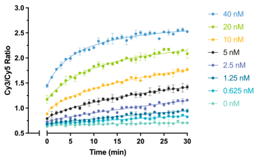
Figure 1: ATP-dependent chromatin remodeling assay

EpiDyne®-FRET Chromatin Remodeling Substrate (EpiCypher 16-4201; 20 nM) incubated with SMARCA2 Remodeling Enzyme (concentrations indicated) in 1x SMARCA Remodeling Assay Buffer. Curves denote FRET efficiency/chromatin remodeling.

Epicypher热销产品——SMARCA2 Chromatin Remodeling Enzyme (Human BRM)

Figure 2: Protein gel data

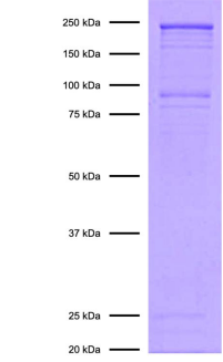
SMARCA2 Remodeling Enzyme (1 µg) was run on an SDS-PAGE gel and stained with Coomassie blue. The migration and molecular weight of the protein standards are indicated.

订购详情

货号

产品名称

规格

15-1015

SMARCA2 Chromatin Remodeling Enzyme (Human BRM)

100 Reactions

如需了解更多详细信息或相关产品,请联系EpiCypher中国代理商-上海金畔生物 

M.O.M(Mouse on Mouse) 免疫检测试剂盒

M.O.M(Mouse on Mouse) 免疫检测试剂盒

M.O.M. 免疫检测系统是专门设计用于小鼠来源一抗检测小鼠组织样本,可显著降低二抗与内源性Ig的非特异性结合,降低背景染色。M.O.M.试剂盒包含Vector Laboratories专利性M.O.M小鼠Ig封闭试剂。M.O.M.试剂盒检测系统有两种形式:一是亲和素-生物素检测系统(包含M.O.M. Elite ABC试剂盒、Fluorescein试剂盒和Basic试剂盒),二是聚合物检测系统(M.O.M. ImmPRESS HRP Polymer试剂盒)。根据多抗原染色操作步骤,M.O.M.免疫检测试剂盒可轻松实现两个或多个抗原的标记。可以在同一个组织切片上使用多种小鼠来源的一抗,而无需考虑待检组织样本的种类。使用M.O.M.检测系统可轻松获得高质量染色结果。

● 当使用小鼠来源一抗检测小鼠组织样本时,可显著降低内源性小鼠Ig染色

● 操作简单

● 无需繁琐的计算

● 无需抗体预结合步骤

● 可使目的蛋白呈现出清晰的特异性染色

● 与荧光素、酶等检测系统兼容

● 试剂盒形式多样:可提供含或不含酶(或荧光素)形式

M.O.M(Mouse on Mouse) 免疫检测试剂盒 M.O.M(Mouse on Mouse) 免疫检测试剂盒

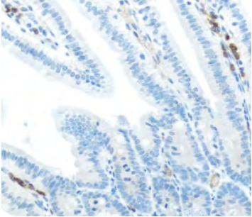
使用标准抗小鼠IgG polymer系统(左)和Vector M.O.M. ImmPRESS HRP Polymer 试剂盒(右)对小鼠肠组织

进行染色。棕色信号是IgG背景染色。(两者均未孵育一抗,使用Vector DAB显色、苏木精复染)

Recommended applications:

Studies in geneticallyengineered mice

Transgenic and knock-out models

Mouse xenograft tissue

Normal mouse tissue

M.O.M(Mouse on Mouse) 免疫检测试剂盒

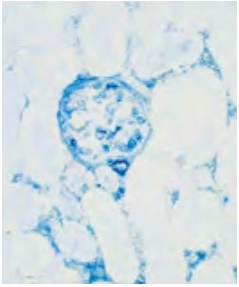
小鼠平滑肌肌动蛋白抗体用于小鼠肾脏组织染色:

使用VECTASTAIN ABC-AP试剂盒和Vector Blue substrate(蓝色底物)显色。采用标准生物素化抗小鼠抗体和正常的封闭血清,可以看到高背景染色(左)。

采用Vector M.O.M. Basic试剂盒,染色结果背景值低,可获得特异性染色(右)。

相关产品信息

产品名称 产品编号(货号)
M.O.M. Peroxidase Kit  PK-2200 
M.O.M. Fluorescein Kit  FMK-2201 
M.O.M. Basic Kit  BMK-2202 
M.O.M. ImmPRESS HRP Polymer Kit  MP-2400 
M.O.M. Mouse Ig Blocking Reagent  MKB-2213 
M.O.M. Biotinylated Anti-Mouse Ig Reagent*  MKB-2225 
M.O.M. ImmPRESS HRP Polymer Anti-Mouse Reagent MPX-2402

*MKB-2225必须与M.O.M.小鼠Ig封闭试剂(MKB-2213)配合使用,不能单独使用。

详情请咨询 Vectorlabs 中国代理- 上海金畔生物科技